Artifacts Are Extracerebral
Artifact on EEG is anything that does not reflect activity of the brain itself, and can arise from physiologic, electric, or environmental factors. Identifying artifact is both challenging and important: it tends to be everywhere, doesn't follow the rules of localization, and is often intermixed with important, real brain activity. Furthermore, some artifact can look similar to cerebral signals, even taking on rhythmic properties that could be mistaken for seizures. With practice, though, it becomes easier to discern the particularities of artifact from actual cerebral activity.
Eye Blinks
Eye blinks are one of the most common artifacts you’ll encounter, marked by high amplitude positive waveforms in the bifrontal regions. They are seen due to Bell’s Phenomenon; the eyes’ cornea is positively charged and retina is negatively charged; when you blink, the eyes roll up slightly, and the positive cornea moves closer to the frontal electrodes Fp1 and Fp2, which thus see a positive signal that is reflected on EEG.
Eye blinks should really only be seen in the frontal leads, without a prominent field into the posterior regions. A similar waveform is seen upon eye closure, and a sort of opposite waveform of a large bifrontal negative charge is seen upon eye opening. Eye blinks are a key component of a normal awake EEG. Be careful not to mistake them for frontal / anterior predominant spike and waves.
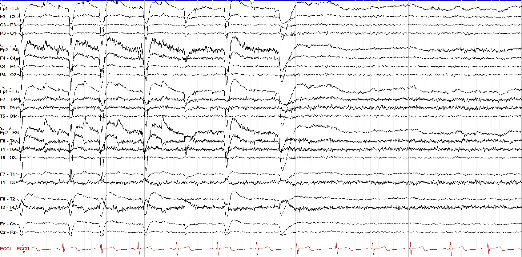
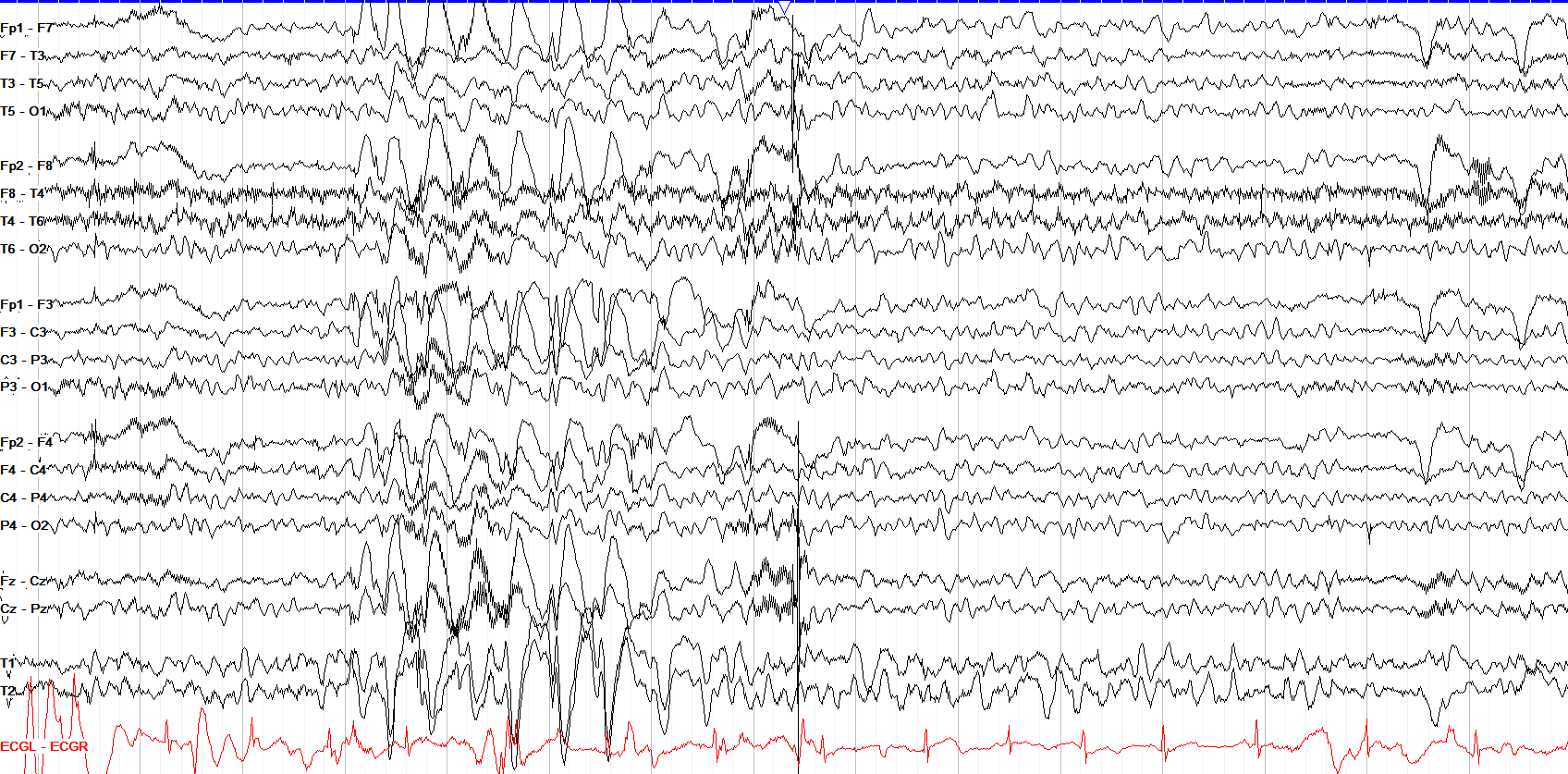
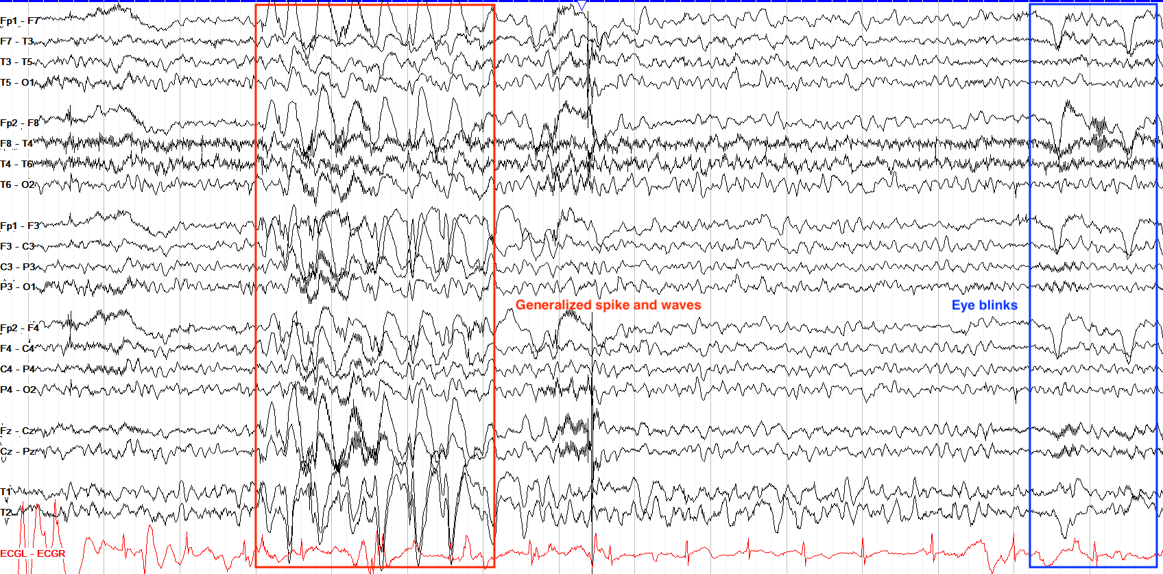
This tracing shows multiple high amplitude generalized spike and waves with an anterior predominance, which are notable for a broad field that goes all the back to the occipital regions. However, toward the end of the page we also see a few eye blinks. Note how different they look to the generalized discharges—first, they have no field posteriorly; second, they have no preceding spike before the larger amplitude wave, and third, they have no disruption of the background.
Lateral Eye Movements
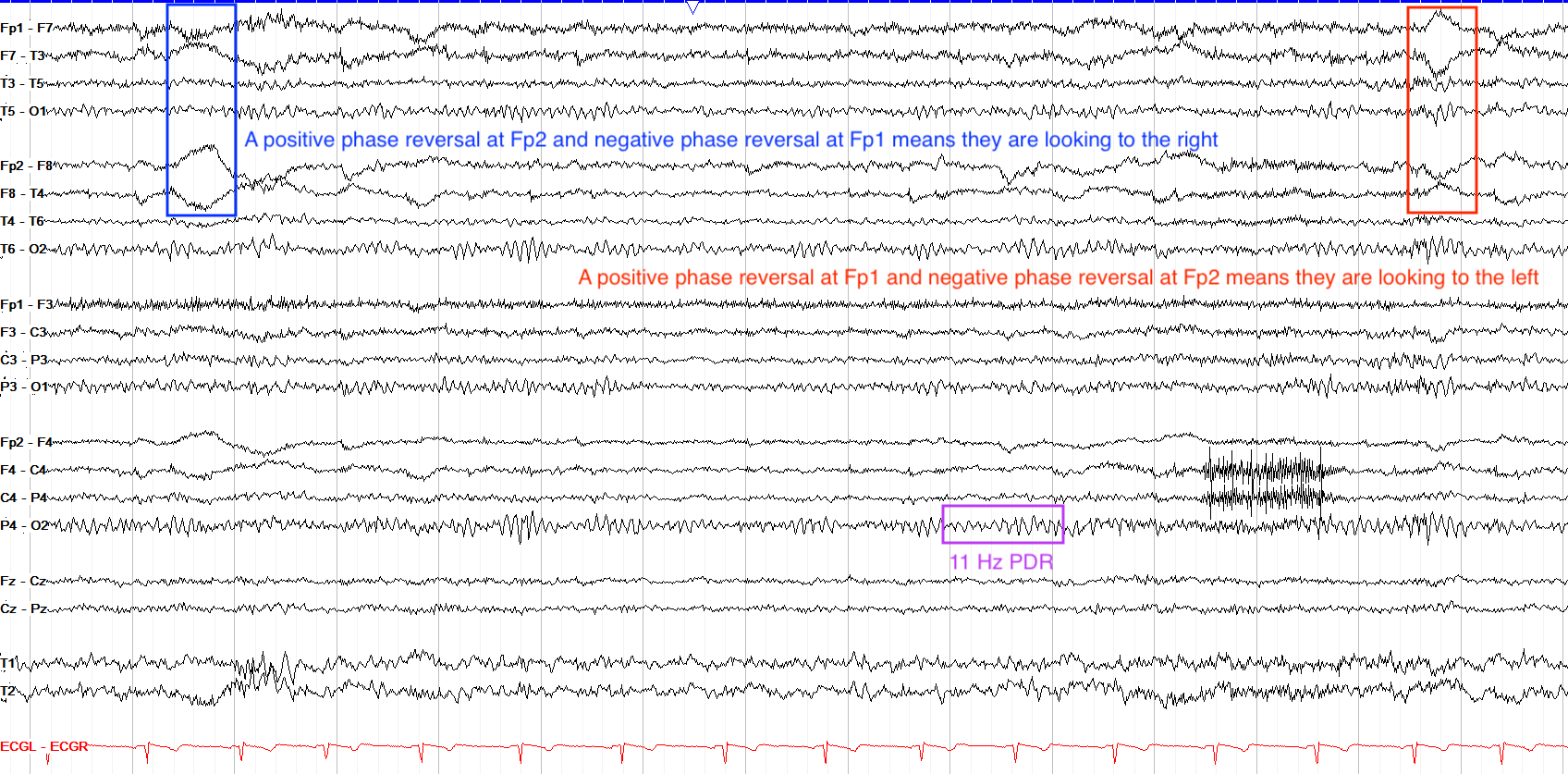
Lateral eye movements are marked by opposing polarities in the F7 and F8 leads. This is due, again, to the positive charge of the cornea and negative charge of the retina. When you look to the right, the right cornea gets closer to the F8 electrode, which sees a positive charge; the left retina gets closer to the F7 electrode, which thus sees a negative charge. So, with lateral eye movements you look to the positive side.
On a bipolar montage, this produces a phase reversal: because F8 becomes the point of maximal positivity when looking right, its neighboring channels open away from it (a positive phase reversal), with the mirror-image negative phase reversal at F7. See the montages chapter for the full breakdown of how a phase reversal’s polarity differs from a single deflection’s.
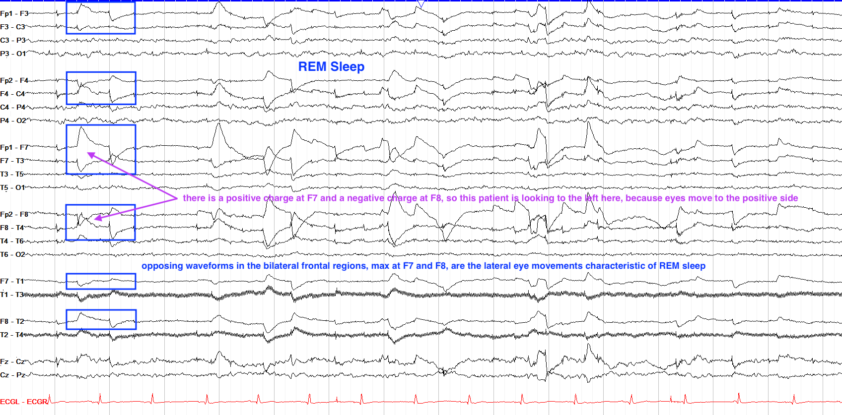
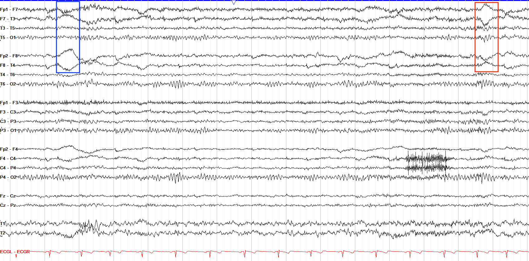
Recall that EEG helps you keep a positive attitude, so you always look to the positive side on EEG. As such, when considering F7 and F8—the electrodes maximally affected by lateral eye movements—when you see a positive phase reversal (the leads move away from one another) in the F8 leads, the patient is looking to the right, and should have a complementary, simultaneous negative phase reversal (the leads move toward one another) at F7. Other notable things on this tracing are a good PDR of around 11 Hz, a normal AP gradient, some myogenic frontal muscle artifact, and likely a drowsy state given the slow somewhat undulating frontal eye movement artifact in the temporal chains.
Chewing & Hypoglossal
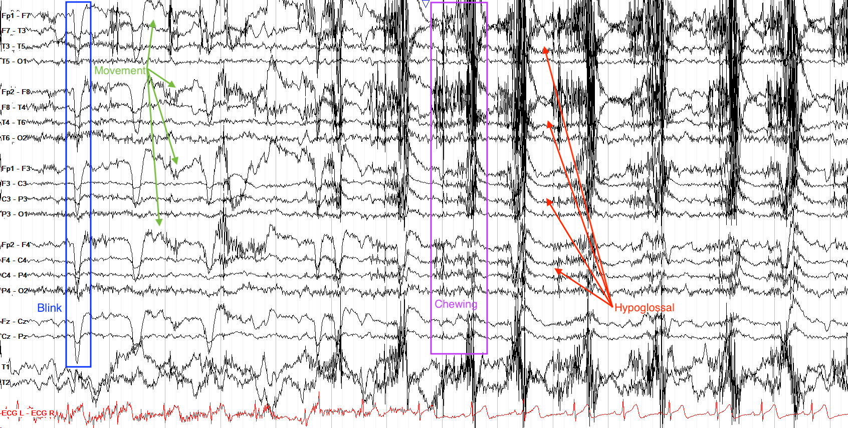
Chewing and tongue (glossokinetic, or hypoglossal) artifact are rather hard to miss when they crop up. Chewing artifact is really just muscle artifact from the temporalis muscle, and is marked by sudden onset, intermittent bursts of generalized very fast activity (muscle artifact). It’s easy to characterize with video correlate, as you can just look at the video to see if they’re chewing, but even without video chewing artifact does not usually share a close morphology with any other important physiologic activity. Just be careful not to mix it up with generalized paroxysmal fast activity, which is an epileptiform pattern that tends to be slightly slower (beta frequency) and lower in amplitude.
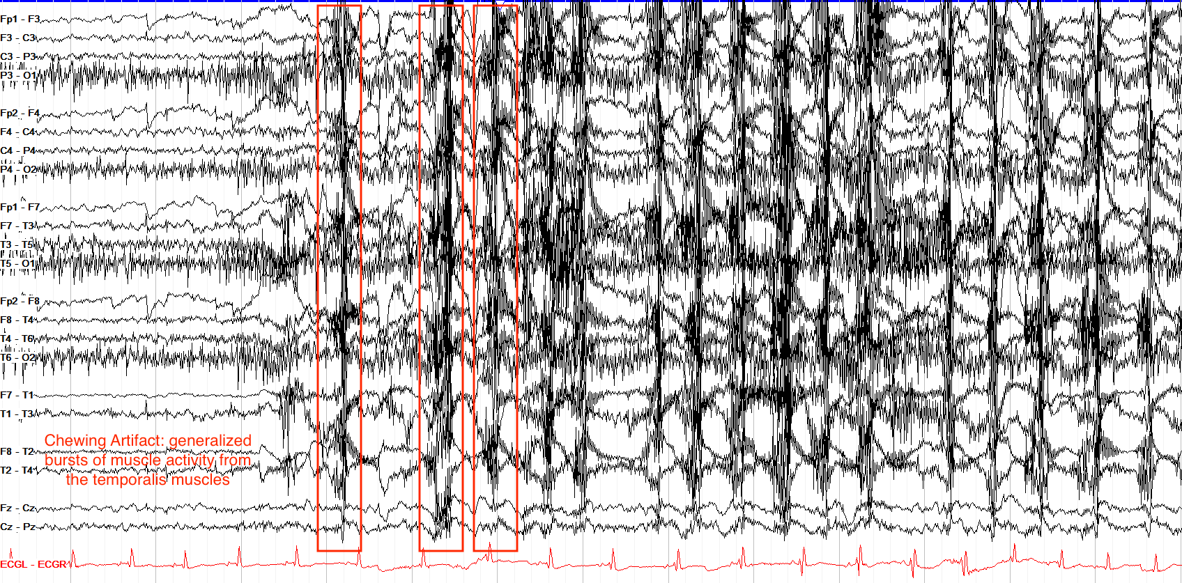
Hypoglossal artifact is often but not always seen with chewing artifact, and appears as slower, diffuse delta frequency activity from the movement of the tongue. Notice how, on the example below, all the tracings have essentially the same movement—this is too organized for something like slow wave sleep, and while ictal patterns can lead to such rhythmicity, there is no evidence on this single page of the evolution across time or location that characterize seizures. One notable point about tongue movement artifact is that it is reproducible—you can ask the patient to move it, or to say “la la la” (aka a lingual phoneme) to see if the same pattern is brought out on the EEG.
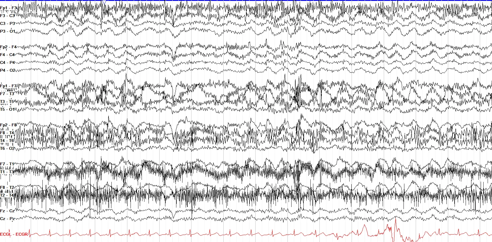
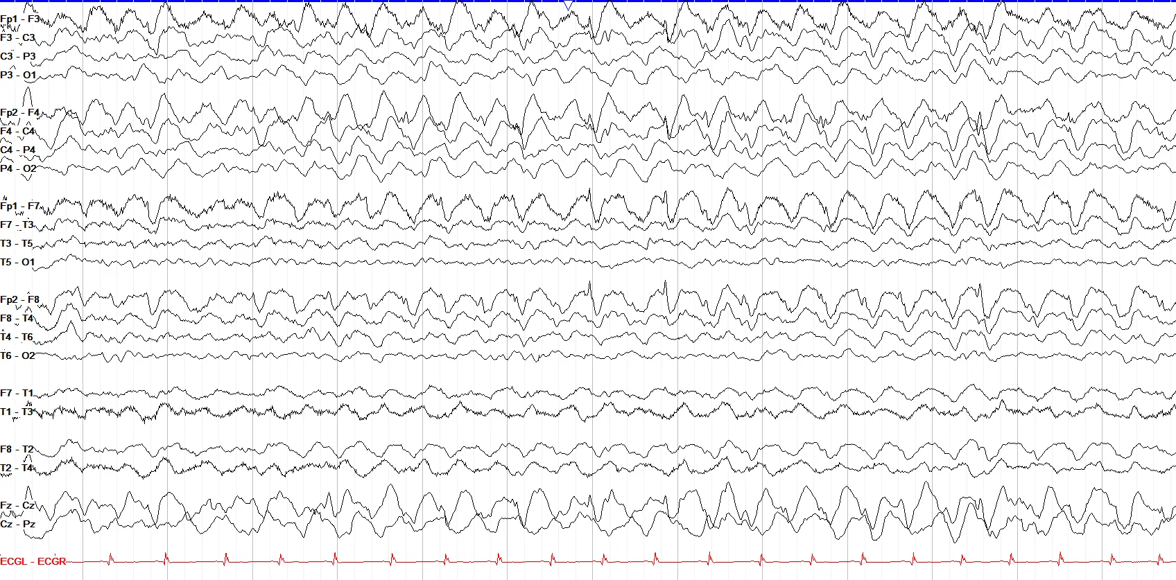
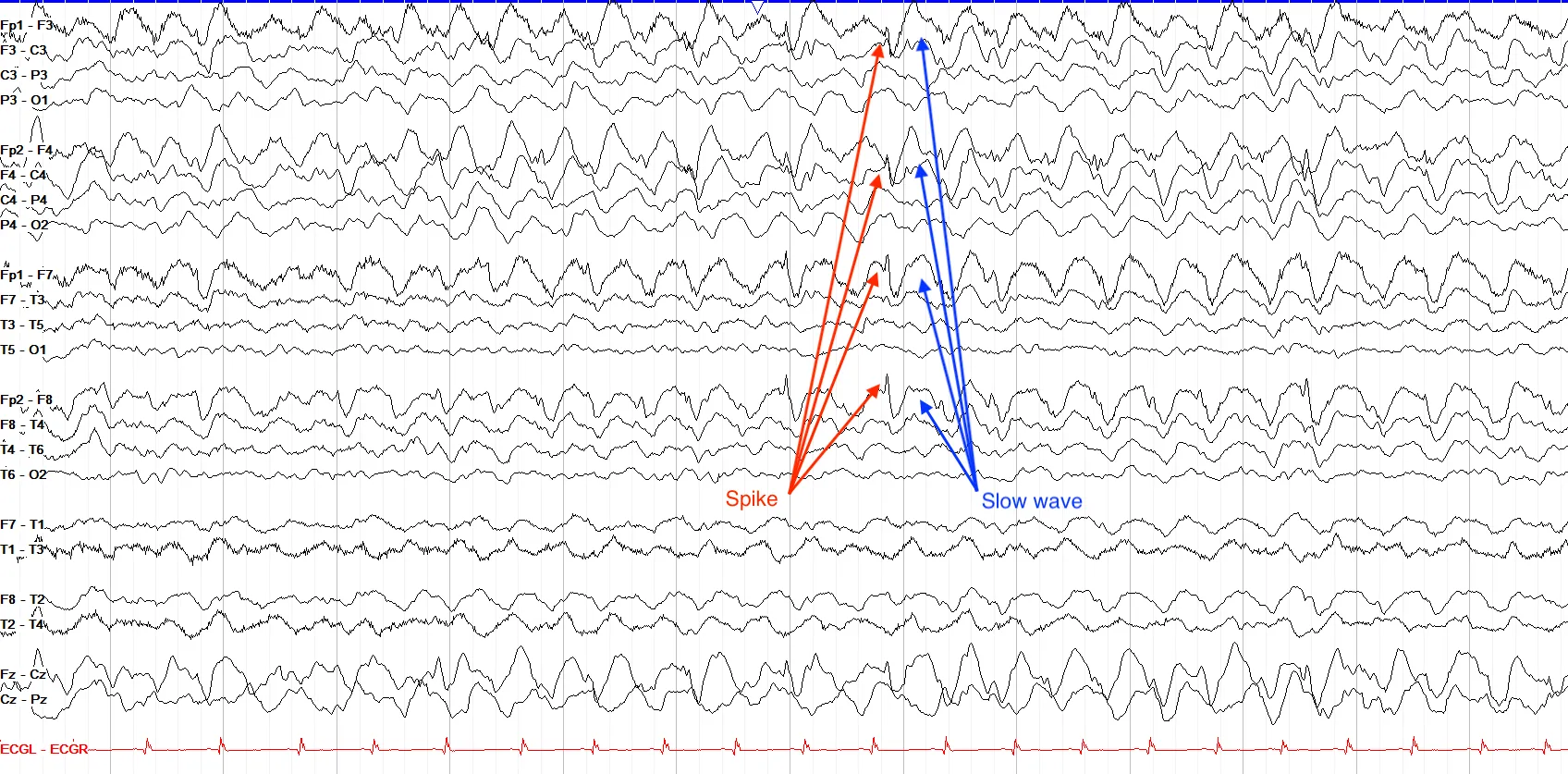
Don’t mistake this tracing for hypoglossal artifact! This page shows rhythmic 2.5 Hz bifrontal spike and wave activity. Notice first that the waves are not quite generalized, as they taper off into the occipital region in the temporal chains (hypoglossal artifact tends to be more generalized). Second, and even more importantly, notice the morphology—each waveform is composed of a small spike and an aftergoing slow wave, which is not typical for hypoglossal artifact either.
ECG & Cardiac
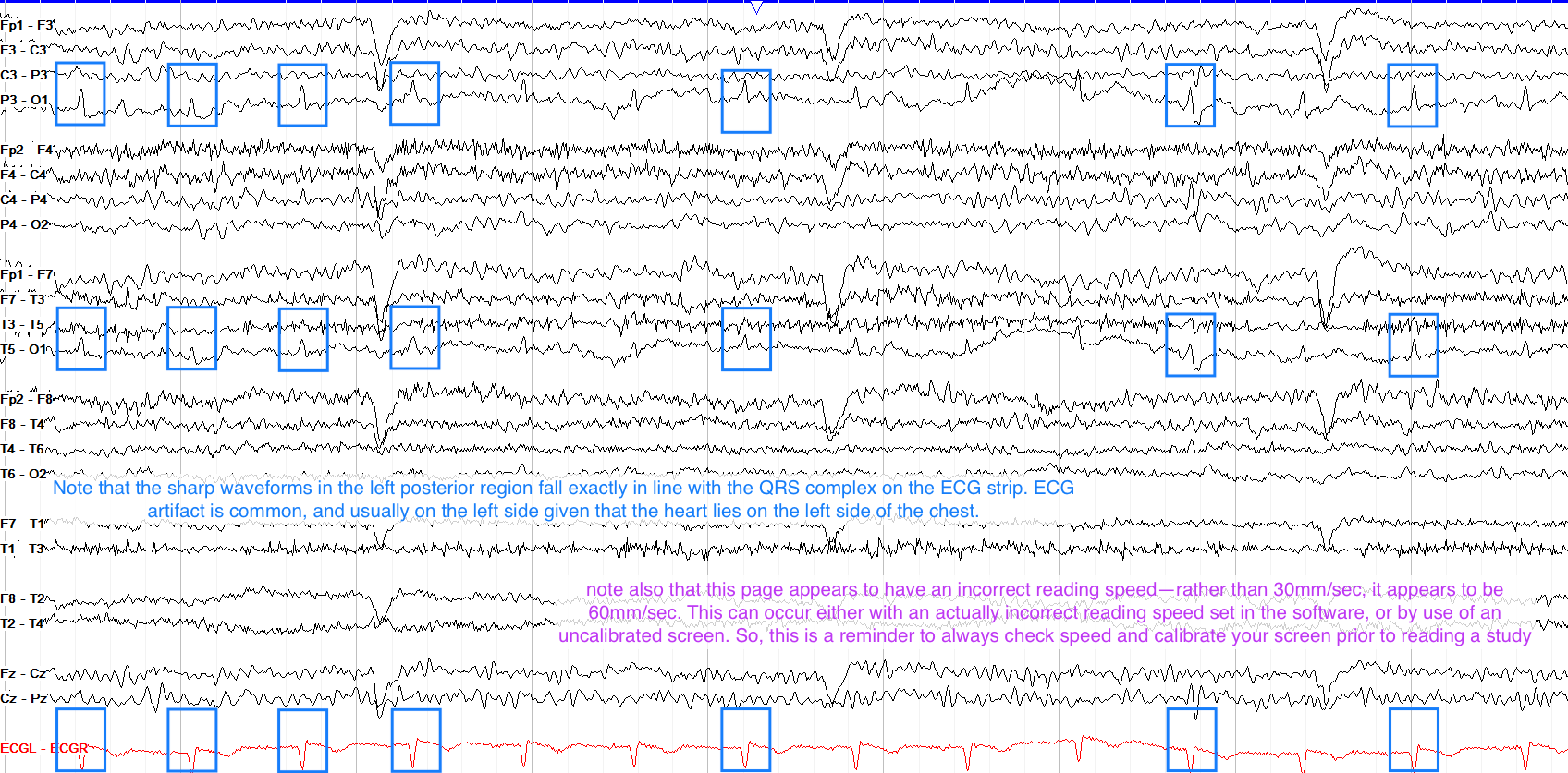
ECG artifact is marked by waveforms that are time locked to the QRS complex on the ECG tracing. They tend to be more prominent or solely seen on the left side, because the heart is in the left half of the chest. While they’re usually relatively low amplitude, as the example below shows they can be more prominent but should not be mistaken for posterior epileptiform discharges or POSTS (neither of which would be time locked to the QRS).
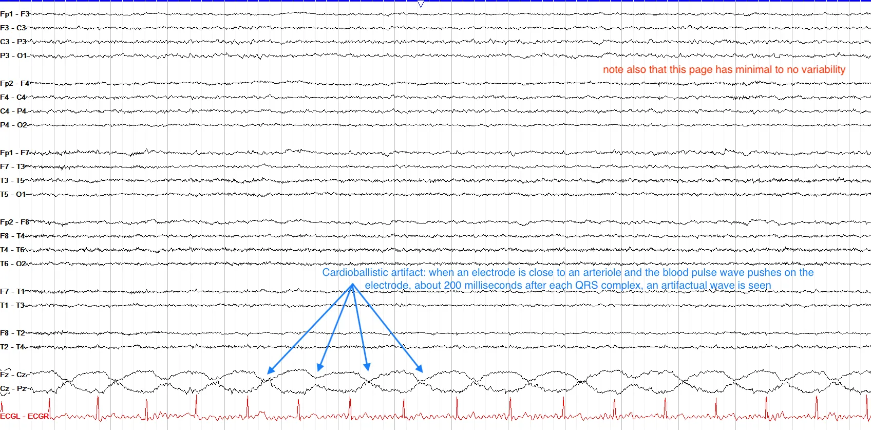
A much less commonly seen cardiac artifact is cardioballistic artifact, in which the EEG electrode is placed just above an artery, and each pulsation of the artery is picked up as motion artifact on the EEG.
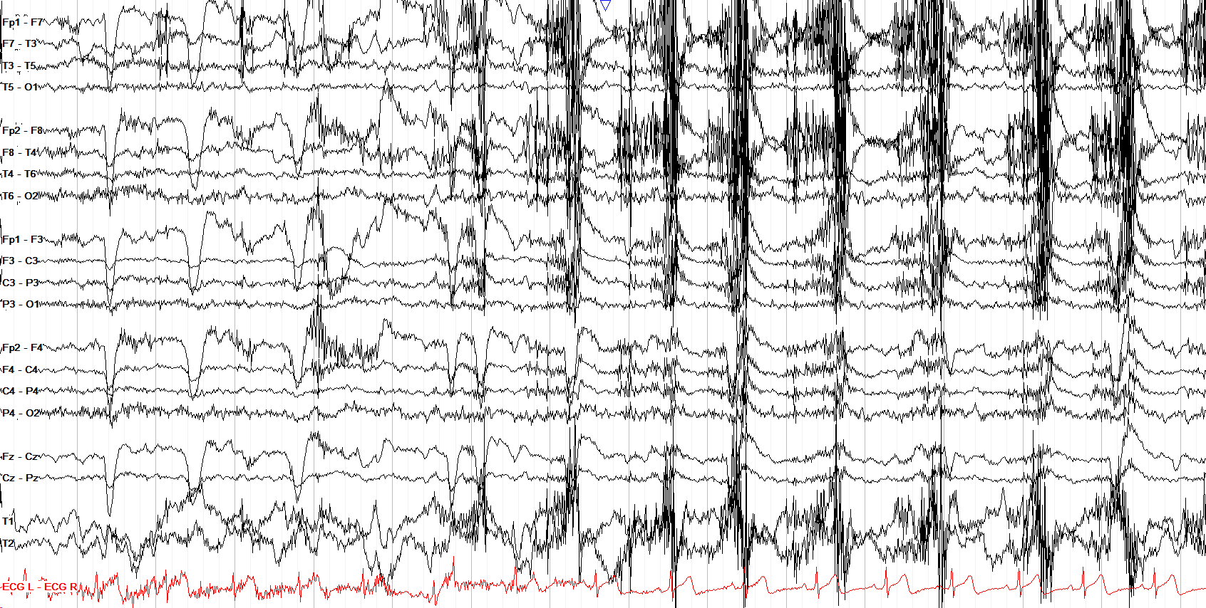
This page from a normal tracing shows many artifact types typical for the awake state. Most prominent are the early eye blinks and the aggressive chewing in the back half of the page with some hypoglossal movement seen in between the bursts of myogenic artifact from the chewing. Of note, there is also myogenic artifact elsewhere, most prominent frontally, and there are higher amplitude, disorganized movement artifacts also seen best frontally. Not marked on the page are a few lateral eye movement artifacts interspersed among everything else.
Electrical & Electrode
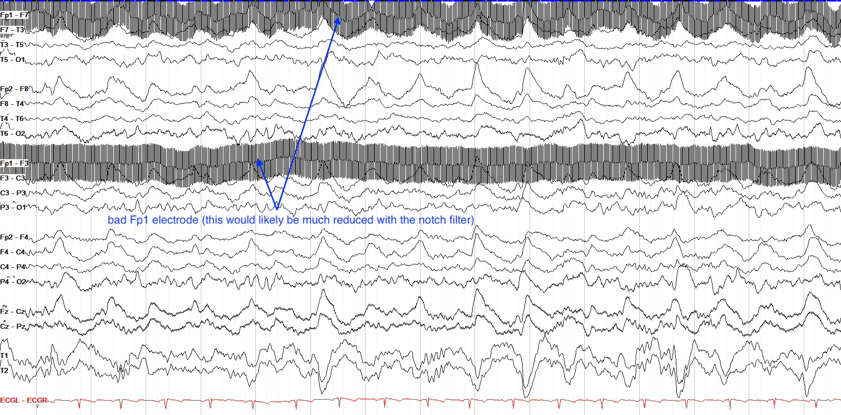
Electrical artifact stems from the interference of surrounding equipment, suboptimal grounding, and more; it can be caused by anything from an electrical appliance to a cell phone charging. It appears as a very fast, very monotonous activity that runs at 60 Hz in the USA and 50 Hz in Europe and elsewhere, based on characteristics of local electric grids. Most modern EEG equipment is quite good at minimizing this artifact type, and you can further use the notch filter to selectively remove all the activity at 60 Hz or 50 Hz (because no cerebral activity that fast is reliably detected by scalp EEG, this usually doesn’t affect interpretation; a notch filter can still subtly distort sharp transients, though, so it’s applied judiciously). In the example below, the notch filter is not on, and the Fp1 electrode is likely not well connected, leading to a lot of electrical interference.
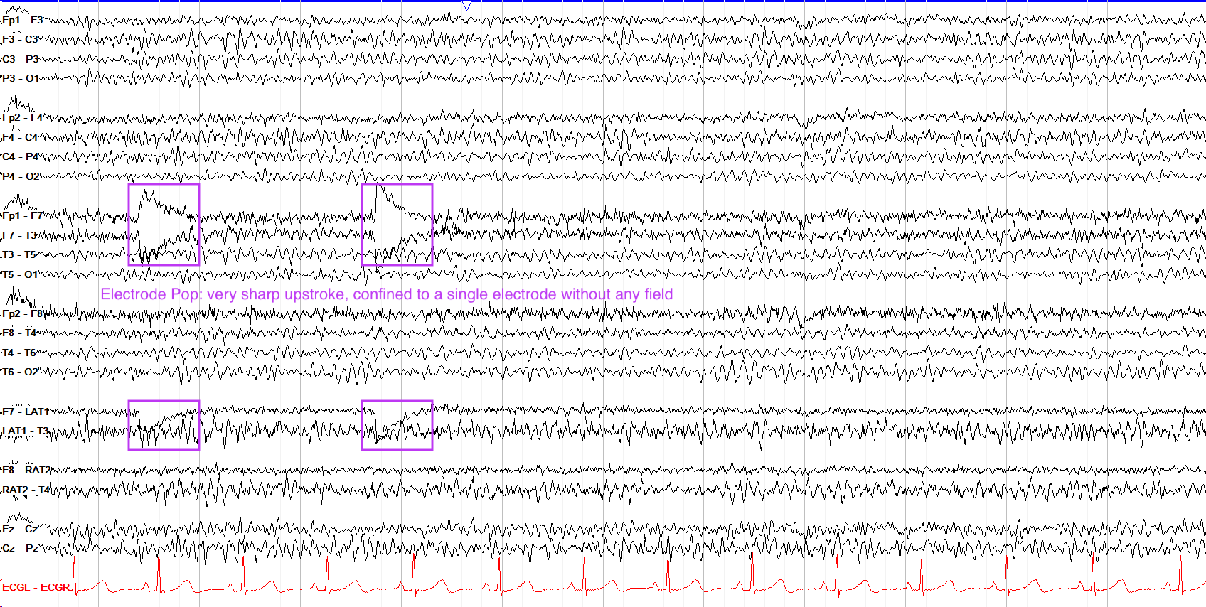
Electrodes themselves can give artifact as well, usually in the setting of them becoming loose over time or getting bumped by things in the environment. A common such finding is electrode pop, caused most often by a loose electrode. On EEG pop is marked by a single electrode showing a very sudden, steep upslope with a slower downslope and absolutely no field. On the example below, F7 is involved; if this continues, you should take a look at the electrode to make sure it’s not falling off.
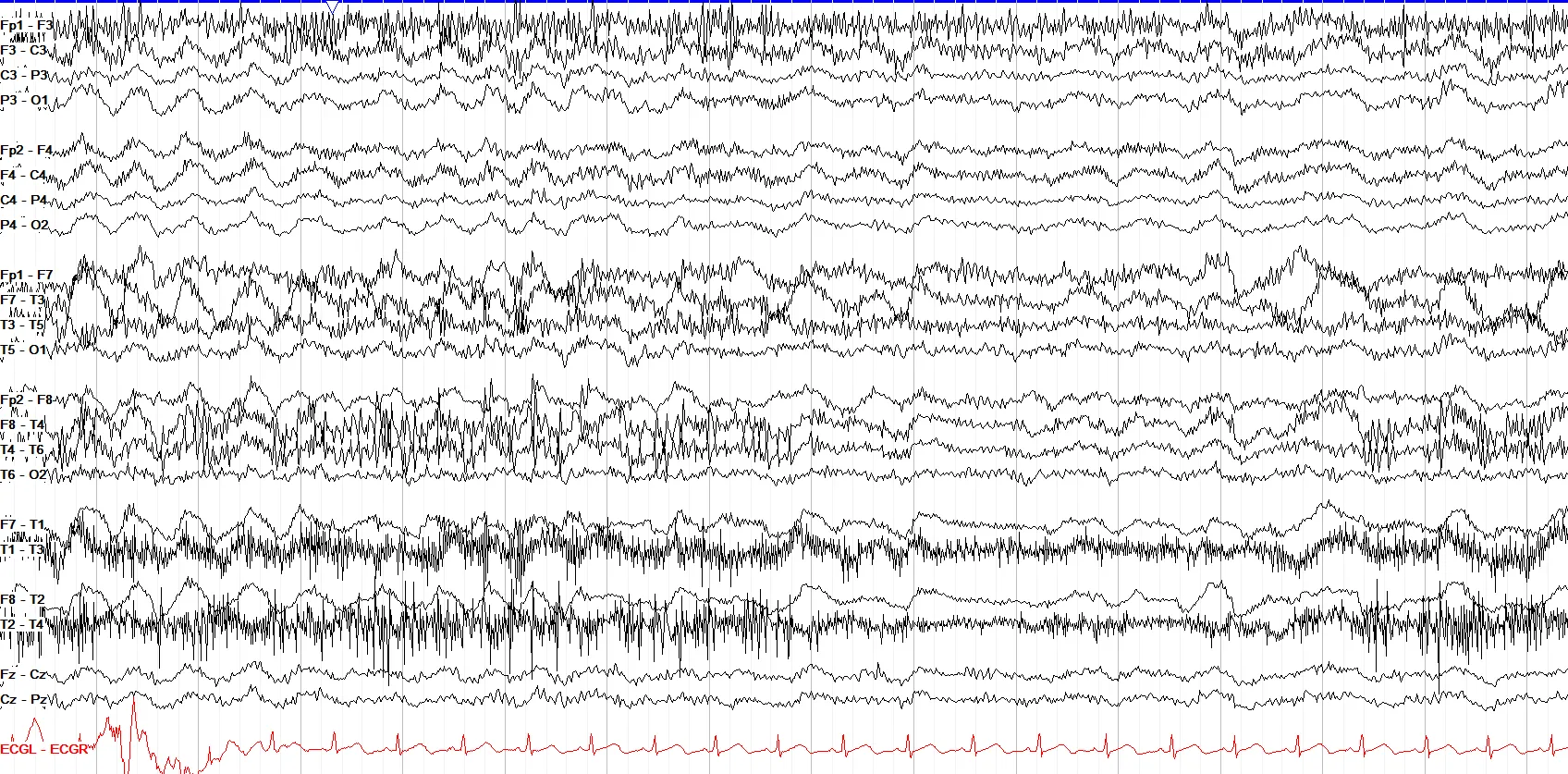
This tracing shows prominent hypoglossal artifact, marked by diffuse slow activity. There is also a lot of myogenic artifact from the lateral temporal leads, likely from the temporalis muscle. Towards the end of the page (the last three seconds), there is some movement artifact seen as “sloppy” appearing and disorganized slow activity slightly more prominent frontally and in the temporal chains.
Myogenic & Muscle
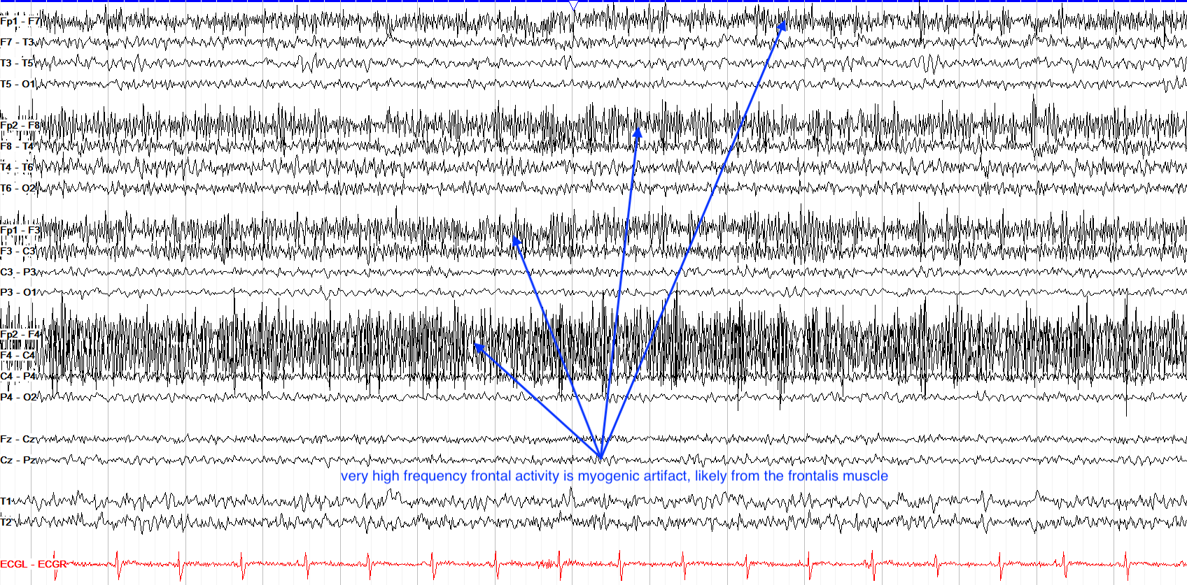
Myogenic artifact comes from muscle movements, and is most commonly found in the frontal or lateral temporal regions due to tension or movement of the frontalis and temporalis muscles. It is marked by high frequency, often low amplitude activity overlying the normal cerebral rhythms, and is usually most prominent in the awake state. Of note, there’s typically only minimal myogenic artifact near the vertex so if you see fast activity there, be slightly more suspicious (although, realistically, myogenic activity is much faster than the cerebral activity that can be picked up on scalp EEG).
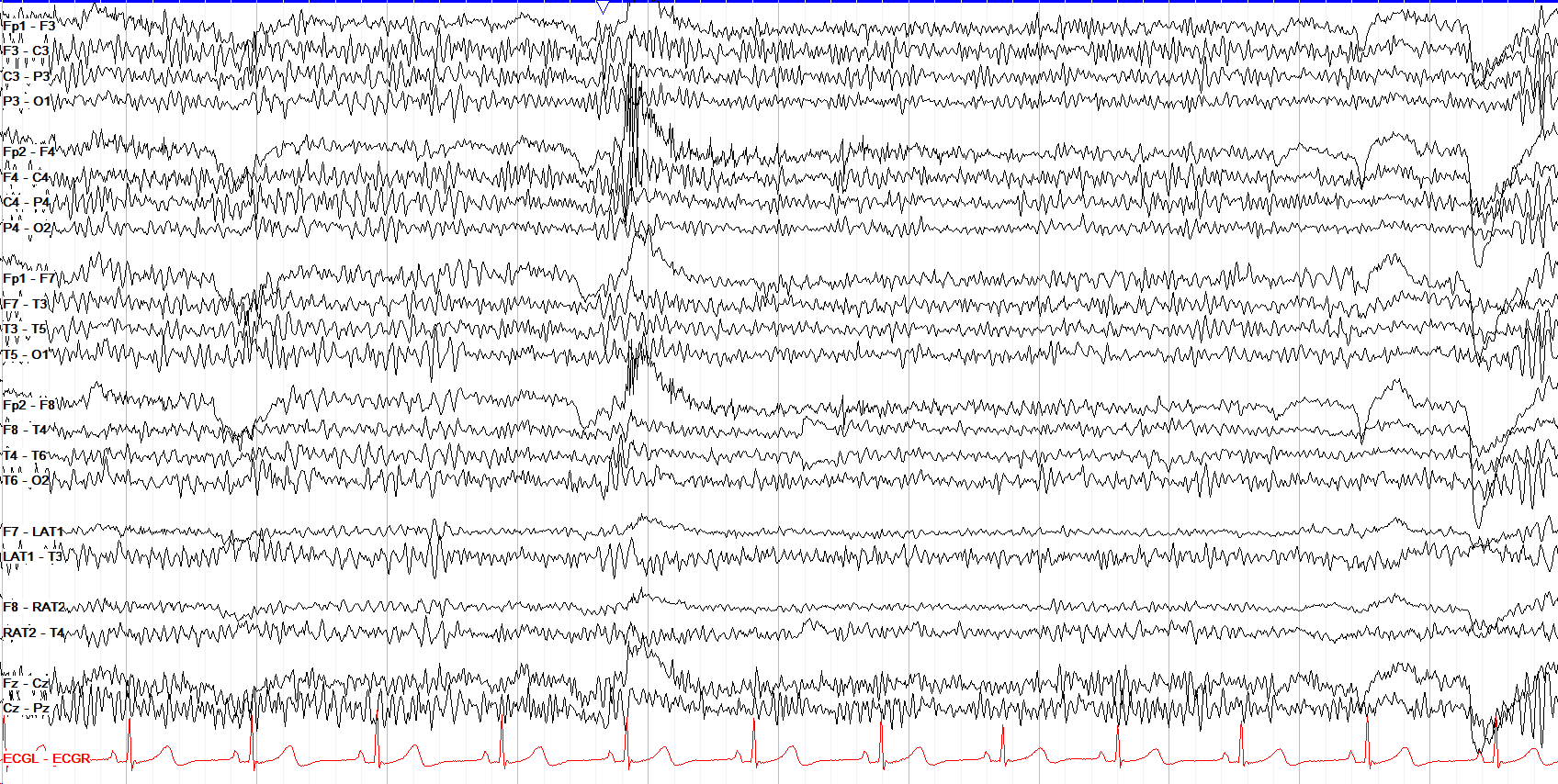
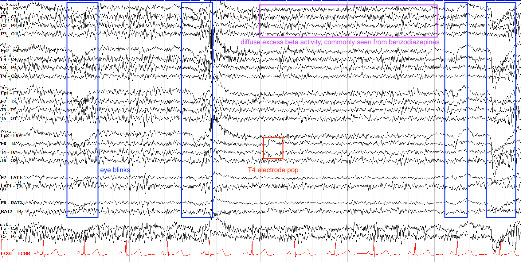
This page has four eye blinks, seen as high amplitude frontal positive deflections. More pertinent, however, is something perhaps less apparent to early learners: diffuse excess beta activity. Notice all the low amplitude fast activity overlying the activity in all the leads—this is excess beta. It is most commonly seen in the setting of benzodiazepine use, and is considered an abnormal background finding. Do not mistake this excess beta for myogenic activity, which tends to be even faster in frequency. There is also an “electrode pop” at T4, marked by a sudden discharge with very steep upslope in a single electrode without any field; this is just a sign of perhaps the electrode being touched, or some other physical issue (if these are persistent, take a look at the electrode to ensure its firmly in place).
Sweat & Other Artifacts
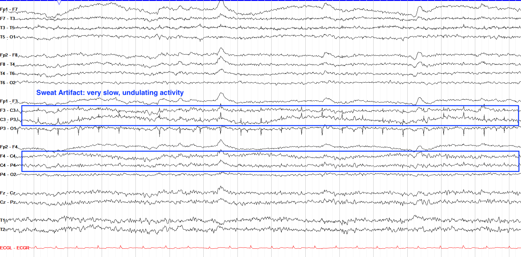
Sweat artifact is marked by very slow (typically less than 0.5 Hz), relatively low amplitude activity that arises because the sodium chloride in sweat carries a charge, which is picked up by the EEG electrodes. Sweat artifact doesn’t have to follow a particular pattern in terms of localization, and can be bilateral, unilateral, or even focal to just a few electrodes. The example below also has electrode artifact at P3.
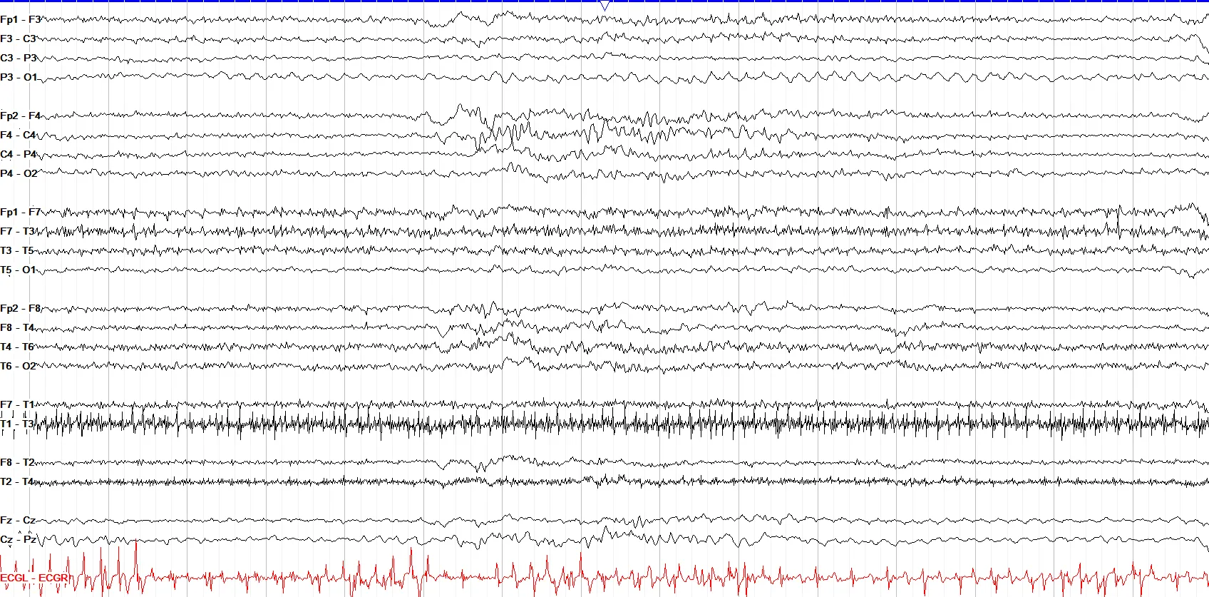
In the hospital, particularly in patients intubated and sedated in the ICU, you’ll commonly come across chest physiotherapy artifact. This is important to know about because it can appear rhythmic similar to a seizure, although you can easily distinguish the two (even without your keen electrophysiologic skills) by looking at the video, if available. On the EEG, it also typically differs from a seizure by its lack of a field or evolution. Chest PT tends to be more prominent posteriorly, but depending on the patient’s position and where the PT is being applied, that can vary widely.
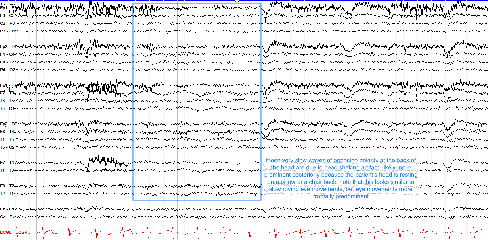
Movement artifact has a plethora of appearances, usually of chaotic looking, high amplitude activity that doesn’t mimic any actual cerebral patterns. However, head shaking artifact can be slightly trickier and is marked by slow, low amplitude activity that is usually more prominent posteriorly if the patient is resting their head on a pillow. It can look similar to the roving eye movements of drowsiness, but eye movements are in the anterior leads.
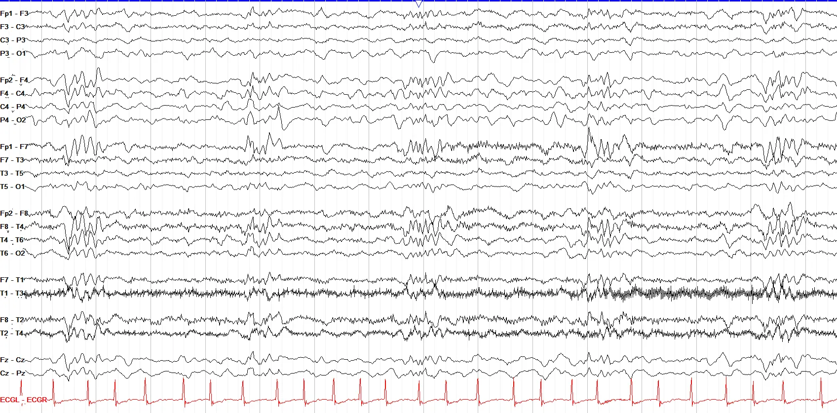
While the above artifacts are perhaps the most commonly seen, you’ll come across many other kinds of artifact as you continue to read EEG. On this example below, for example, in an intubated patient a small bubble on the tip of the nose fluctuated with breathing, leading to a sort of periodic burst of activity that is, in fact, just artifact.
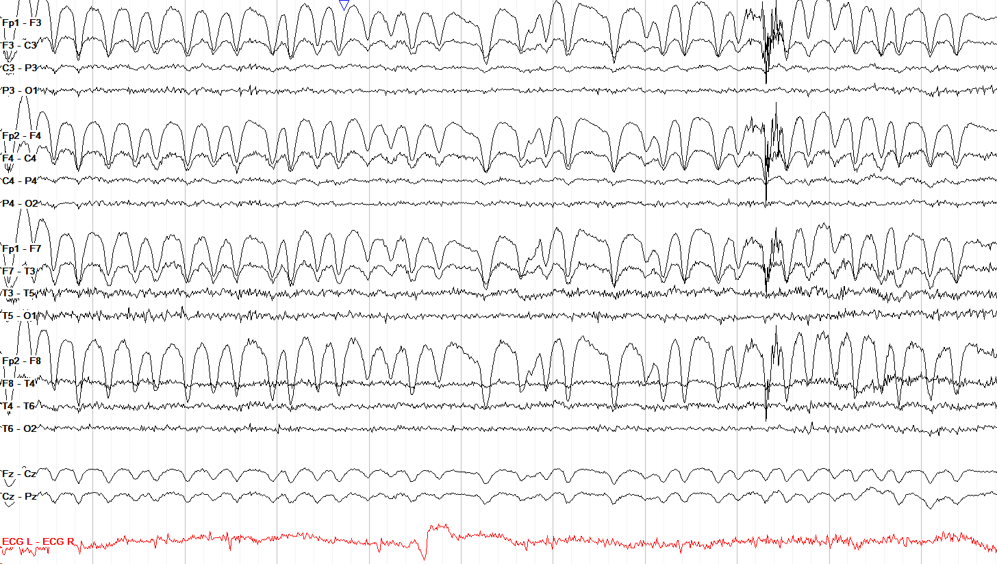
Similar to eye blinks, note that the prominent positive deflections across this page do not extend posteriorly beyond the frontal leads. Given this constricted frontal area involved and the lack of any underlying spike waves to suggest epileptiform activity, these waveforms are most consistent with eye flutter. Perhaps the patient needs some eye drops.
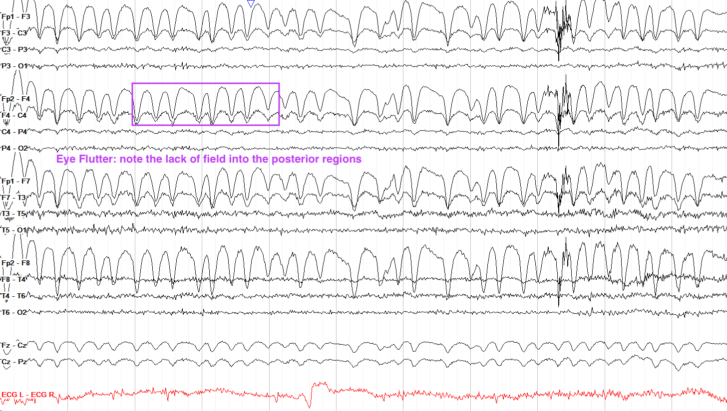
- Eye blinks are large frontal positive deflections due to Bell's Phenomenon. They do not extend to the posterior leads.
- Lateral eye movements show a positive frontal deflection on the side you look toward, and a negative deflection on the opposite side.
- Myogenic/muscle artifact is very high frequency activity, most prominent in frontal and lateral temporal leads.
- Chewing produces diffuse bursts of fast myogenic activity. Hypoglossal artifact produces diffuse, slow, synchronized delta.
- ECG artifact appears as sharp, QRS-locked deflections, usually more prominent on the left side; location and polarity depend on the montage.
- Sweat produces very slow undulating waves due to NaCl charge changes at electrodes.
- Movement artifact is often high amplitude, chaotic, and varies widely depending on the type and location of movement.