The EEG in Epilepsy
Epileptiform activity on EEG is indicative of cortical hyperexcitability, which carries an increased risk for seizures and suggests the presence of an epileptic network within the brain. There are several types of epileptiform activity, including single discharges (sharps and spikes, discussed here) and rhythmic and/or periodic activity (discussed in Rhythmicity, Periodicity & the IIC).
It is important to note that the presence of epileptiform activity is not sufficient for a diagnosis of epilepsy in a patient with no history of seizures; in those who do have such history, epileptiform activity in between seizures (interictal activity) can help localize seizure onset and characterize different epilepsy types. Being able to clearly tell true epileptiform discharges apart from other patterns and normal variants is critical to avoid misdiagnoses based on EEG tracings.
Sharps, Spikes & Slow Waves
An epileptiform discharge is a hypersynchronous, irritable disruption of the usual functioning of the brain, and sharps and spikes are the most classic type. There are six criteria you can use to decide if a particular waveform is a true epileptiform discharge, based on its morphology and the context in which it is seen, per the International Federation of Clinical Neurophysiology (IFCN) and a 2020 follow-up study that found a discharge with five or more of them can be called epileptiform with more than 95% specificity:
- Sharp or spiky shape
- Asymmetry of the up and down slope
- After-going slow wave
- Interruption of the background
- Different duration from surrounding activity
- Anatomically coherent field
Sharp or Spike Morphology
A sharp is a single discharge with a duration of 70-200ms, while a spike is faster with a duration of 20-70ms. While the specific cutoff between them doesn’t seem to suggest one as being more "severe" than the other, the overall shape of them both is important. Sharps and spikes should have a genuinely pointed peak (not blunted or rounded) with a monophasic, biphasic or triphasic shape; recall this means they cross the baseline zero, one, or two times, respectively (but don’t confuse the term triphasic with generalized periodic discharges with triphasic morphology, which are a distinct entity, not overtly epileptiform, and are further discussed in the Rhythmicity, Periodicity & the IIC section). Polyspikes are aptly named, and comprised of multiple contiguous spikes (polyphasic).
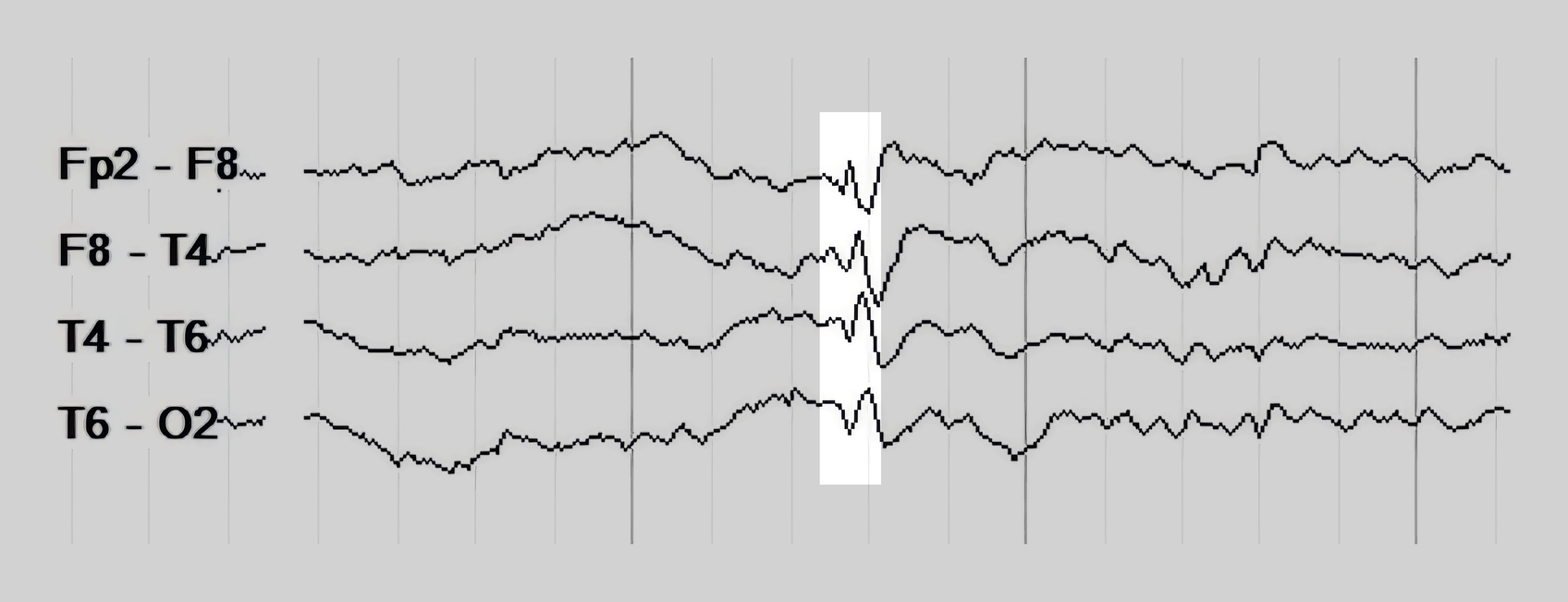
Asymmetry of the Up and Down Slope
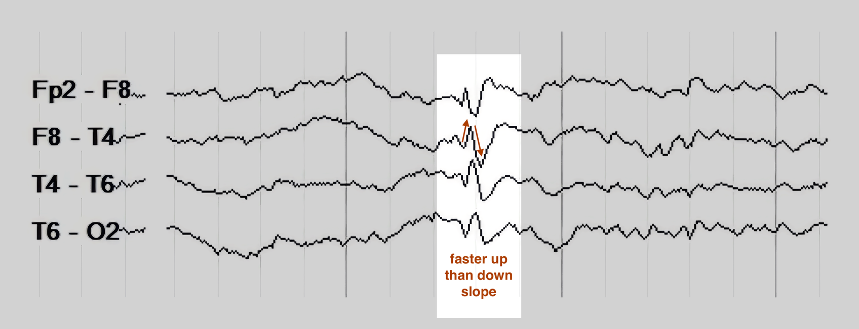
Sharps and spikes tend to have a faster, steeper upslope than downslope, while normal physiological rhythms are usually more symmetric and sinusoidal in their shape. This is because sharps and spikes are reflective of abrupt depolarization of neuronal groups (the upslope) followed by a slower repolarization phase (the downslope).
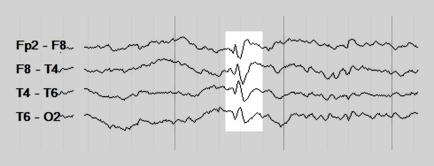
After-going Slow Wave
Some sharps and spikes are followed immediately by an aftergoing slow wave, which may be higher amplitude than the preceding sharp or spike itself. This slow wave represents active inhibition of the irritable neuron population after its large and synchronized EPSPs that led to the spike or sharp itself. The presence of slow wave morphology after spikes and sharps is among the most reliable indicators that a discharge is truly epileptiform (and to push that point further, most normal variants and background activity will not show slow waves afterward).
Interruption of the Background
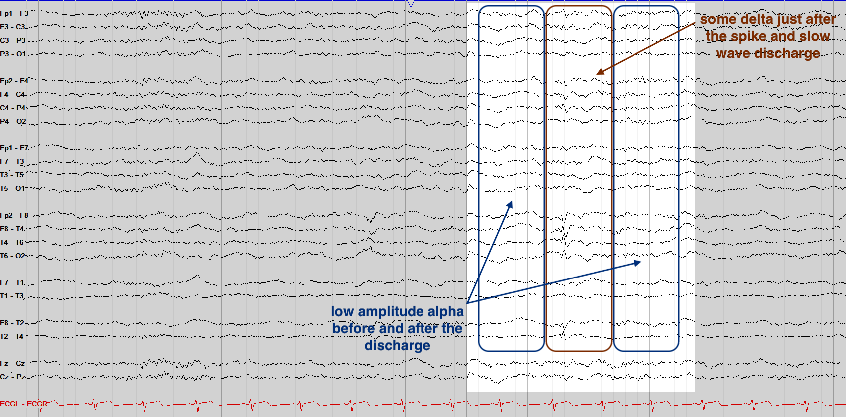
Whether an interictal discharge has an appreciable slow wave or not, it often (but not always) disrupts the background, with subtle slowing, attenuation, or superimposed fast activity on the usual background immediately before or after the discharge. This is, like the slow wave, reflective of the discharge interrupting the usual function of its surrounding area.
Different Duration than Surrounding Activity
True epileptiform discharges should stand out from the background in terms of their frequency / duration. If there is one sharply contoured alpha wave amongst a broader alpha PDR, for instance, that is much less likely to be a true sharp or spike than a higher amplitude theta discharge amongst a background of beta and alpha. You don’t want to mistake a sharply contoured cycle of a normal background rhythm for a discrete epileptiform discharge.
Anatomically Coherent Field
Considering that even a single discharge involves a large group of neurons, true epileptiform discharges should show a field; this is to say, you often see "ripples" of a discharge in the surrounding EEG electrodes, in a distribution that makes sense based on the discharge’s maximal point of voltage. A left posterior temporal discharge, for instance, may have a field into the left parietal and occipital regions, but less likely a separate, simultaneous interruption in the right frontal region. The right temporal discharge below, for example, has a good field throughout the broader right frontocentrotemporal region, while the left side is largely unaffected. As with most of the criteria here discussed, not all discharges will have an obvious field, and multifocal discharge patterns can give multiple fields.
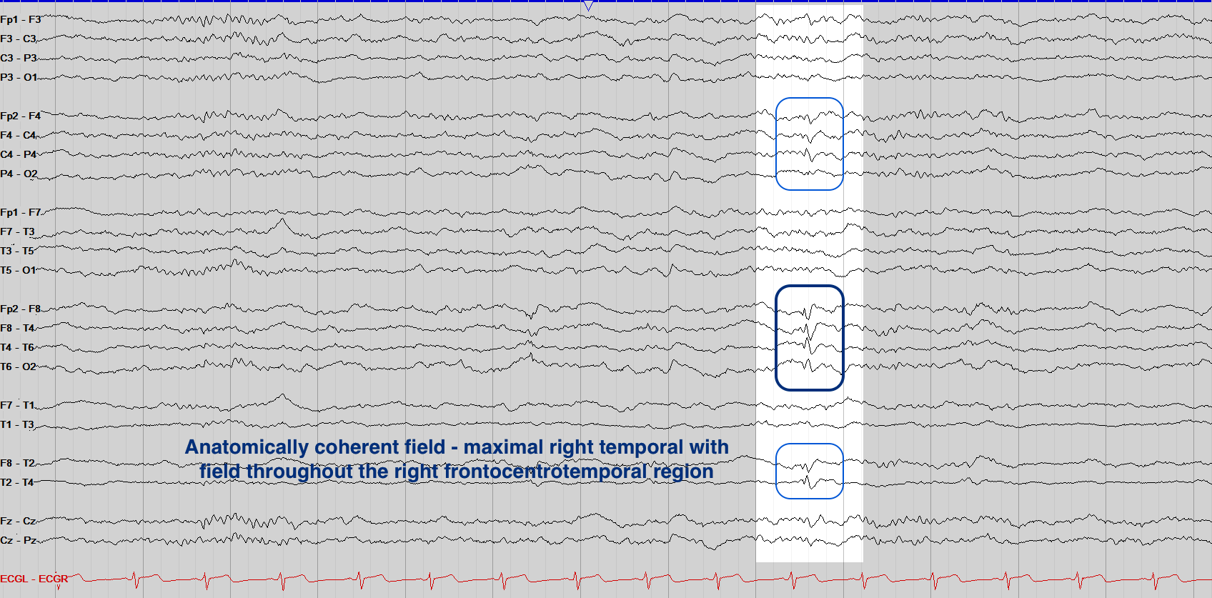
Of these criteria, the three most reliable are pointed morphology, an after-going slow wave, and a clear field. If a discharge has this trio of characteristics, or five or more of them overall, it is almost certainly epileptiform. Discharges that show four overall are still likely epileptiform, but for those with only two or three you want to make note of them, but not call definitively epileptiform unless you see further, stronger examples elsewhere in the recording. While this system for identifying interictal discharges may sound complicated, it is truly a helpful paradigm for accurate reading, and with time you’ll likely run through this list in your head quickly and automatically as you read.
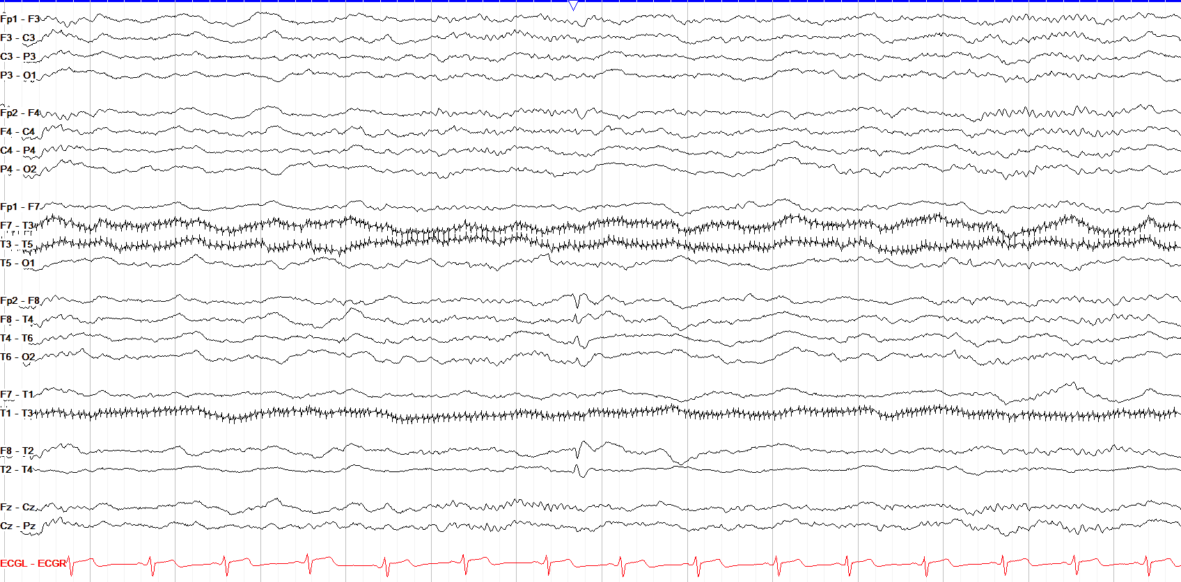
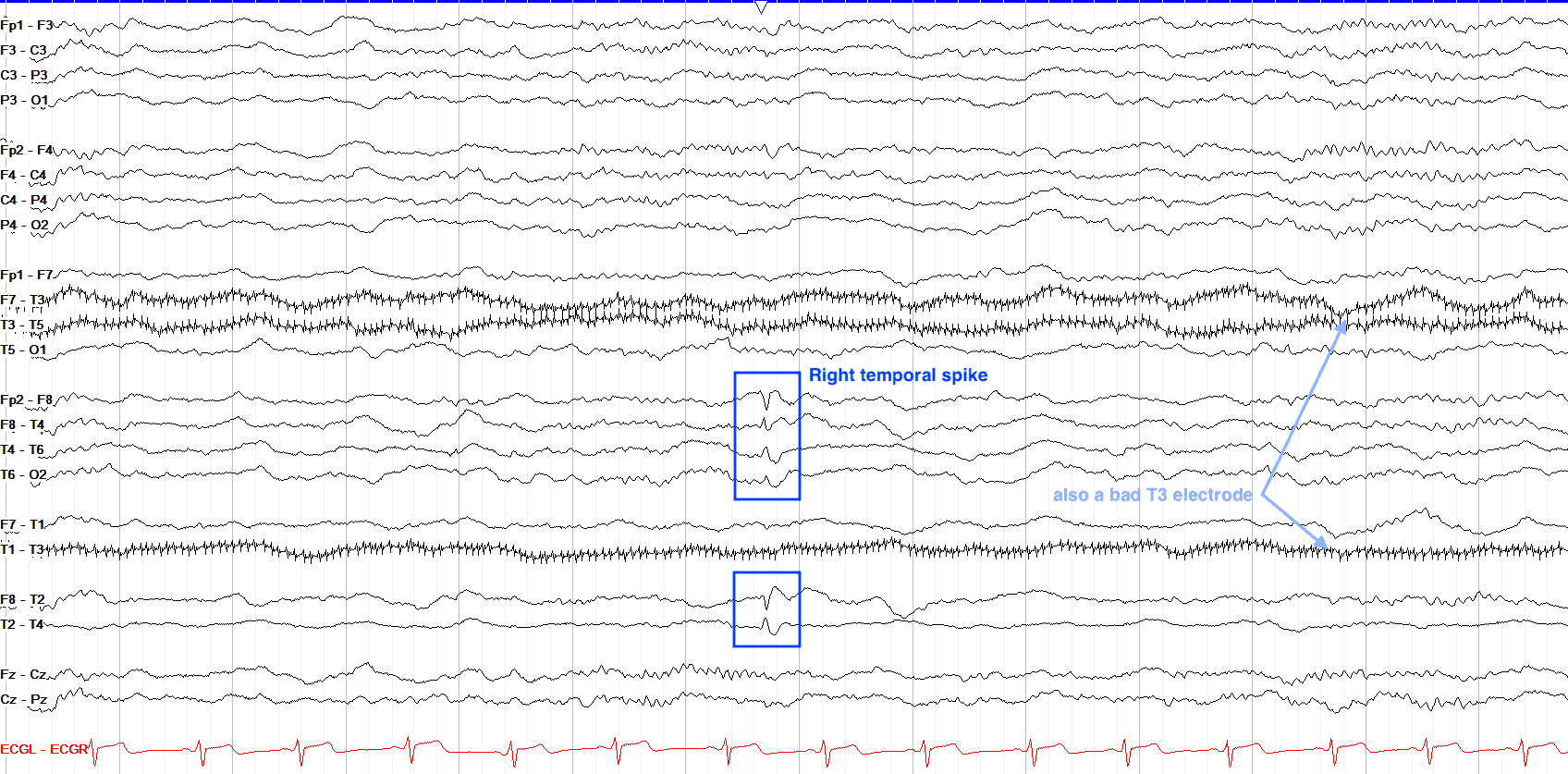
This bipolar tracing shows a phase reversing spike and slow wave in the right anterior temporal region. Note the phase reversal (the point at which two tracings "point to one another") between Fp2-F8 and F8-T4, but also at F8-T2 and T2-T4. This puts the discharge between F8 and T2, in the anterior temporal region. Other notable findings on this tracing are a bad T3 electrode, seen as repetitive fast artifact over the T3 leads, and several low amplitude sleep spindles suggesting this is in stage II sleep.
Localizing the Discharge
Generally speaking, the location of an epileptiform / interictal discharge suggests cortical irritability of that region. For example, a discharge at T4 suggests right mid-temporal hyperexcitability with epileptogenic potential. Bipolar montages are especially helpful in finding epileptiform discharges because they allow the discharges to form a phase reversal, wherein the adjacent channels involving the discharge point to (if negative) or away from (if positive) the electrode of maximal voltage for the discharge.
However, there are a few fine details and caveats to this rule. First of all, generalized discharges cannot be localized. Typically found with generalized epilepsies, generalized discharges begin in such a widespread fashion that they effectively involve the entire cortex simultaneously. It is possible that deeper structures, such as the thalamus, may be involved with generalized discharges.
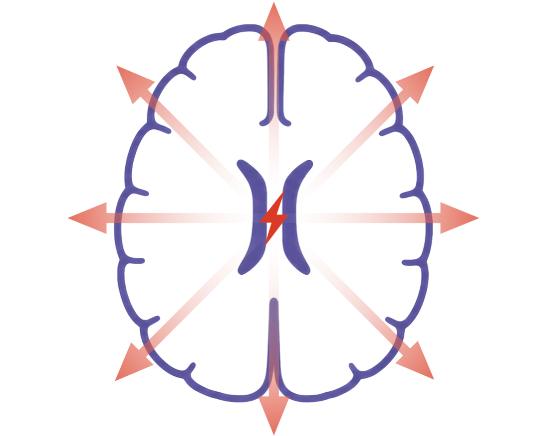
Note that on EEG generalized discharges do not have to be completely the same in every single lead; there is often an anterior predominance to them, for example, but as long as the morphology of the discharge remains throughout all the leads (even if some are less well formed or lower amplitude) and the time of onset is the same in all the leads, you should consider it a generalized discharge.
You may also see discharges that appear generalized but are actually focal with rapid bisynchrony, in which they actually arise from a single location but the networks involved propagate the signal too quickly to trace that location, and they appear generalized. Sometimes with rapid bisynchrony, with careful review you can find a small precedent change before the discharge, such as very low amplitude fast activity, to suggest a lateralized onset.
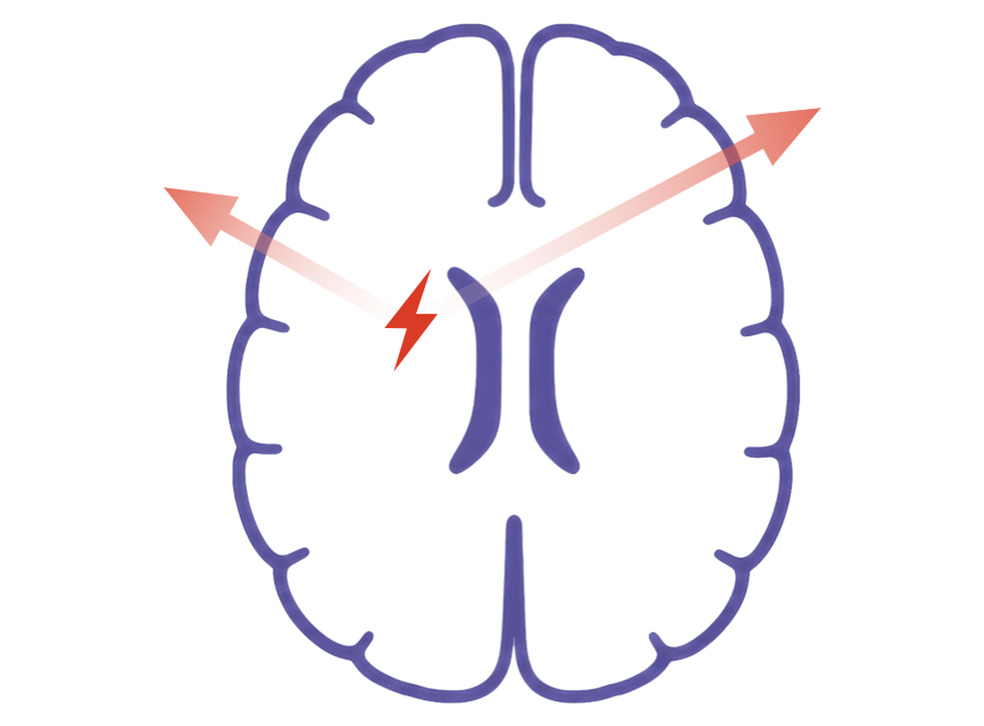
The frontal lobes can be a confusing place for interictal discharges. Deep frontal discharges can be missed on scalp EEG entirely, and mesial frontal discharges can appear as if they are from the contralateral frontal lobe due to the direction of the discharges’ dipole (see below).
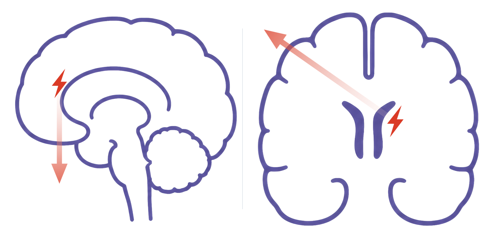
Furthermore, while anterior temporal interictals are perhaps the most common focal epileptiform discharges, because of the proximity of the F7 and F8 electrodes to the inferior frontal region, what appear to be anterior temporal discharges can, at times, actually be from the inferior frontal region; if your montage includes the T1 and T2 electrodes they can be helpful to clarify this.
The central region is less complicated: discharges here are usually normal in the asleep state (i.e. vertex waves from the central region) but not in the awake state. This doesn’t mean that central epileptiform discharges can’t exist, but they should follow the same morphological rules that we discussed above. Furthermore, very mesial discharges from the frontal or parietal lobes can sometimes be seen on the midline EEG electrodes, so if you see midline discharges in the asleep state that are not as symmetric as vertex waves, or with an atypical formation or field for vertex waves, you should be suspicious.
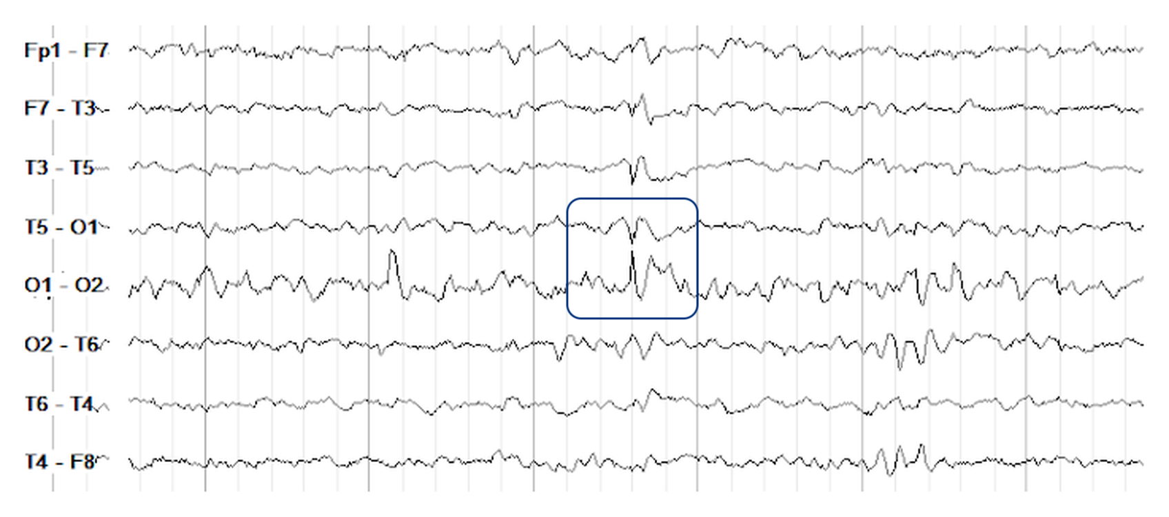
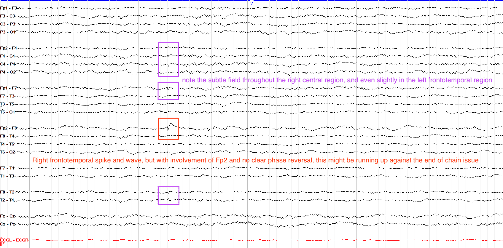
In the occipital region, remember that the O1 and O2 electrodes, similar to Fp1 and Fp2, suffer from the end of chain issue in bipolar montages. That is to say, because there is no electrode behind them to compare their voltage to, there is no phase reversal to stand out on EEG. So, if you see a positive discharge in the occipital regions that is not consistent with a lambda wave or POST, check a circumferential or referential montage to clarify if you're seeing a true interictal discharge.
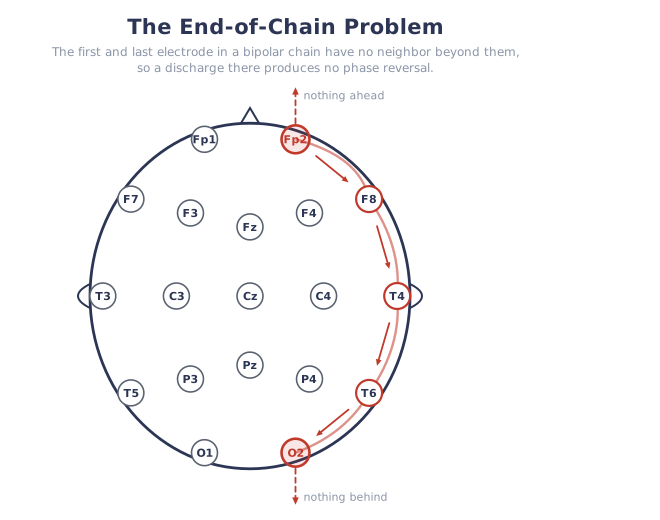
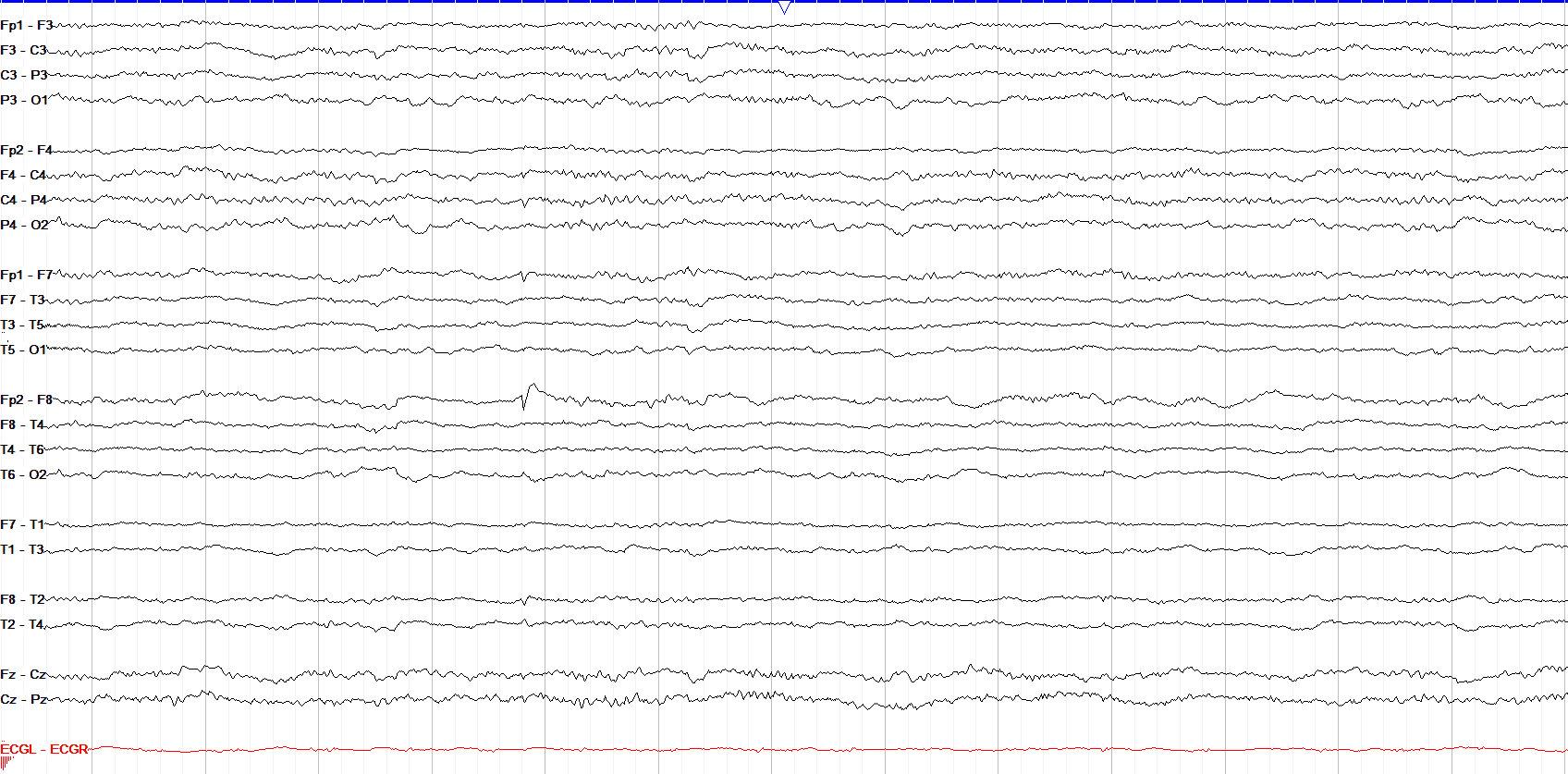
This tracing shows a clear spike and wave discharge at Fp2-F8. However, despite this being a bipolar montage there is no phase reversal. This is a good example of the end of chain issue, where there is nothing anterior to Fp2 to which to compare its voltage. It is possible that the maximal discharge, where the phase reversal would be, is actually anterior to or deep to Fp2, but because there isn't an electrode there to measure it, we can't see the full extent of what would be the phase reversal on scalp EEG. Also note the good field in the right frontocentral region, with some reflection to the left frontal/frontotemporal region.
Paroxysmal Fast Activity
When you see diffuse fast activity, it’s often in the setting of excess beta activity (usually a benzodiazepine effect) and is benign, while very fast activity over the frontal regions is usually muscle artifact. However, paroxysmal runs of fast activity can be epileptiform, and are most commonly seen in patients with generalized epilepsy, Lennox-Gastaut Syndrome, and tonic seizures.
Paroxysmal fast activity can be localized, as in the example below, or generalized. When you see bursts of generalized paroxysmal fast activity, ensure it is not actually a tonic seizure, of which fast activity is classically a key part.
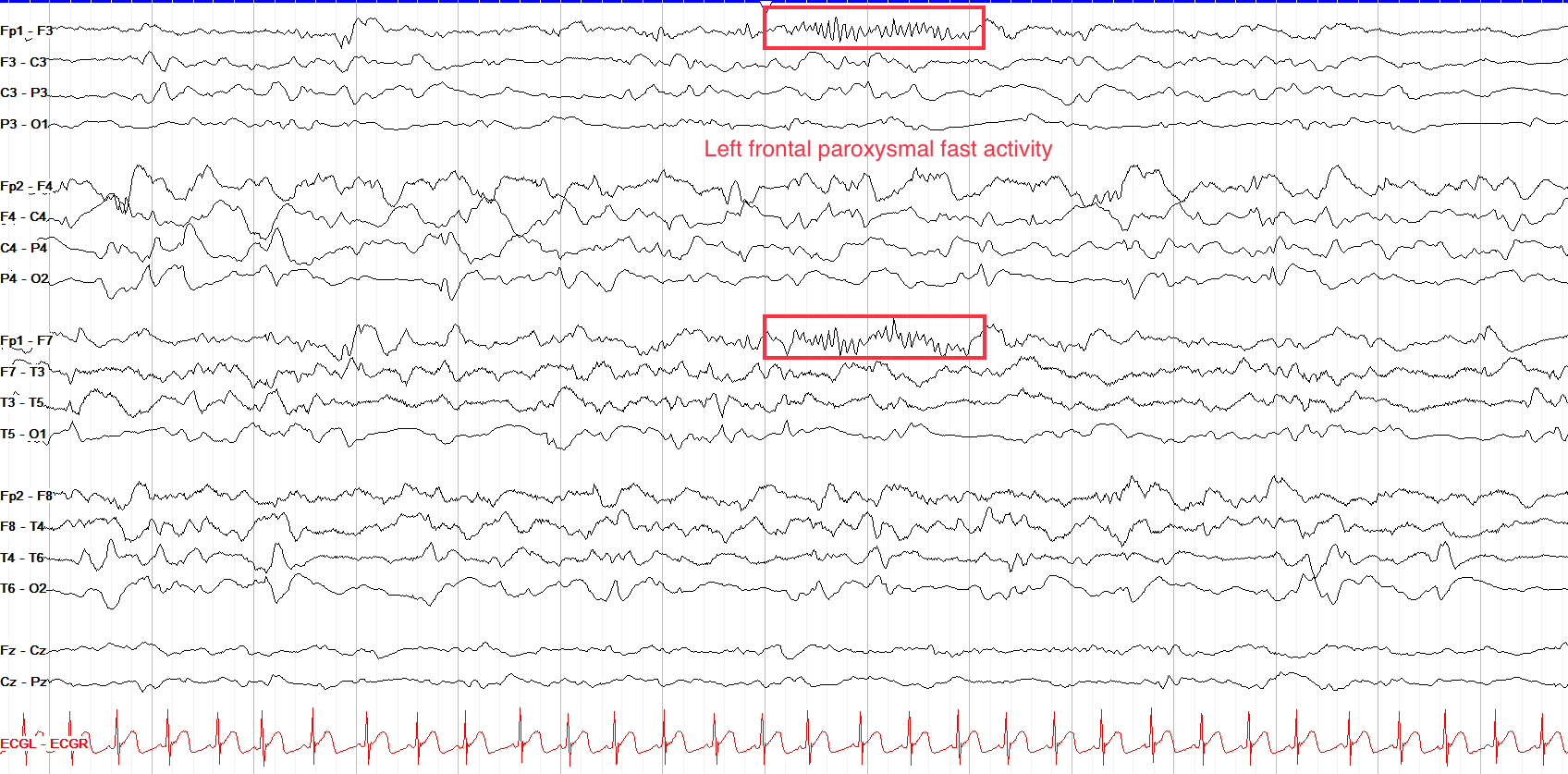
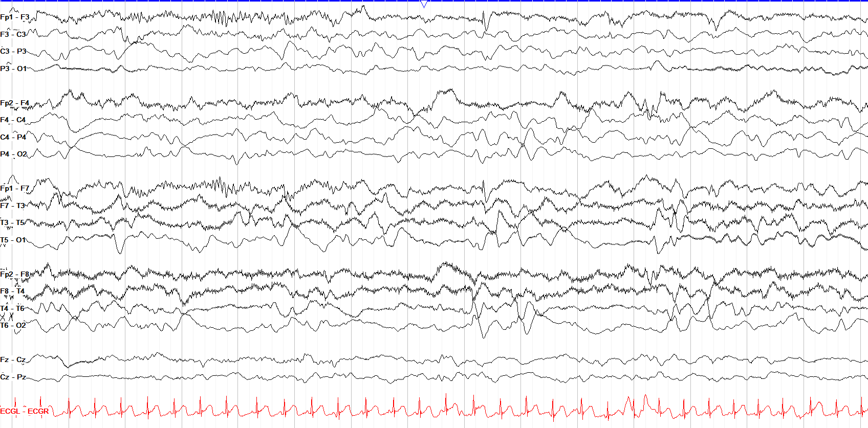
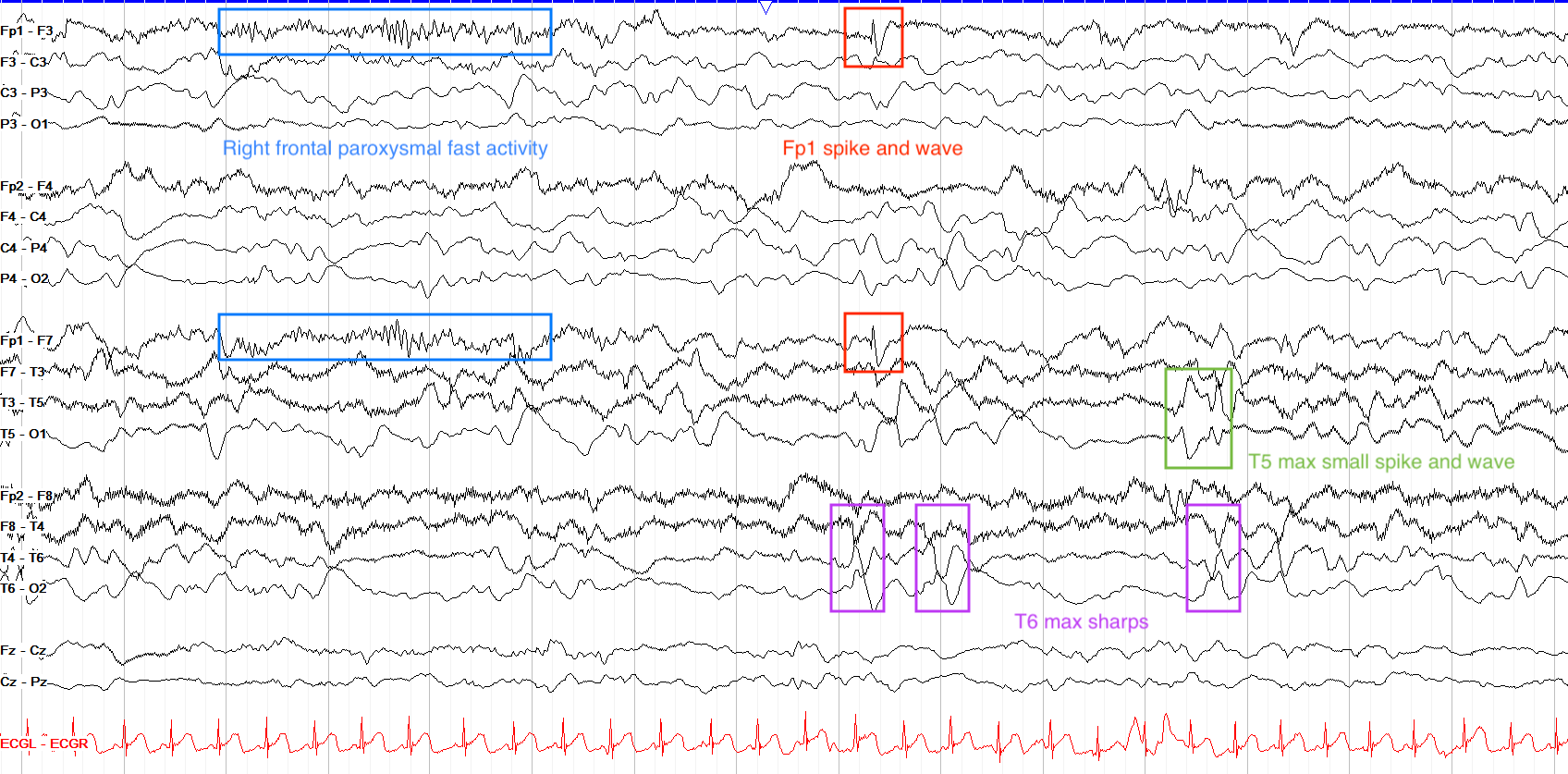
This is a complicated tracing. First note the state of the patient, who appears to be in slow wave sleep (relatively synchronized high amplitude delta activity diffusely, no eye blinks or other evidence of being awake).
There are multiple epileptiform discharges on the page, including a run of left frontal paroxysmal fast activity, left frontal spike and waves (Fp1 max), right mid-temporal sharps (T4 / T6 max, although poorly formed), and left temporal (T5 max) spike and waves. Note the field of the right temporal sharps into the right parasagittal chain. This kind of tracing—disorganized with multifocal discharges—is typical for Lennox-Gastaut Syndrome.
Photoparoxysmal Response
The photoparoxysmal response, while perhaps one of the most well known facets of epilepsy, is in actuality quite rare. It describes the emergence of interictal activity as a result of photic stimulation. Typically, this interictal activity is generalized or focal occipital, arises during but outlasts the photic stimulation itself, and is consistently reproducible at the same frequency of light flashes. Note that photic driving does not confer an increased risk for a photoparoxysmal response or photosensitive seizures.
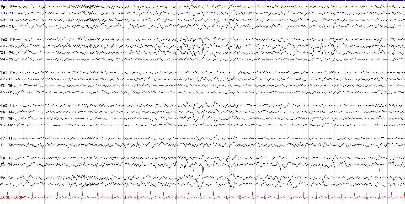
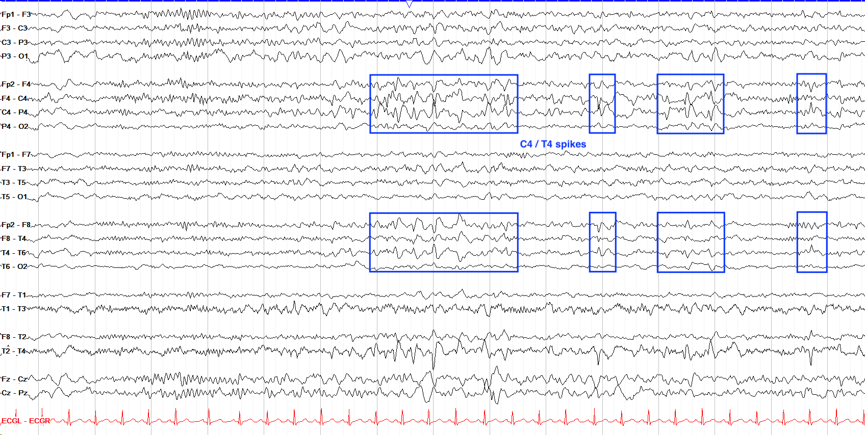
Here we have multiple, very well formed phase reversing spike and slow wave discharges that point toward T4 and C4, with slightly better formed morphology over the temporal than central region. These would thus be right centrotemporal spikes.
Note the field into the left central region, and also that in the right parasagittal chain, the frequent negative phase reversals can appear somewhat similar to electrode artifact, but you don't see evidence of such artifact elsewhere on the page.
- Epileptiform activity on EEG indicates cortical hyperexcitability and increased seizure risk, but its presence alone without clinical seizures does not diagnose epilepsy.
- Spikes and sharps are discrete epileptiform discharges (spikes 20-70ms, sharps 70-200ms) that should show a pointed morphology, after-going slow wave, and anatomical field; less specific traits also include a faster up than down slope, background disruption, and different duration than surrounding rhythms.
- Localization of discharges reflects regional cortical irritability, but beware of generalized vs focal with rapid bisynchrony, mesial frontal “false-localizing” patterns, anterior temporal vs inferior frontal confusion, and end-of-chain issues in occipital regions.
- Paroxysmal fast activity (pFA) is burst-like fast activity in the beta range or faster, often associated with seizure onset or interictal discharge localization; focal pFA more concerning than diffuse, and distinguish from muscle artifact.
- Photoparoxysmal response (PPR) describes the emergence of interictal activity as a result of photic stimulation; it is quite rare, consistently reproducible at the same flash frequency, and photic driving alone does not confer increased risk.
- Always correlate EEG findings with clinical history, age, sleep state, and behavioral context, as many patterns can mimic epileptiform activity and context is essential for accurate interpretation.