The Developing EEG Varies by Age
Newborn babies less than four weeks of age are termed neonates, and their EEGs reflect the rapid development of their brain in that time. The normal neonatal EEG exhibits many characteristics that would be grossly abnormal in an adult, including diffuse slowing, discontinuity, asynchrony, and minimal reactivity, but all of these characteristics should organize over standard increments of time into the familiar tracings of infants, children and adults. It seems that babies like to keep both their parents and neurophysiologists on their toes.
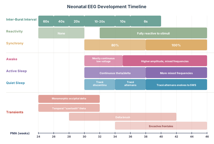
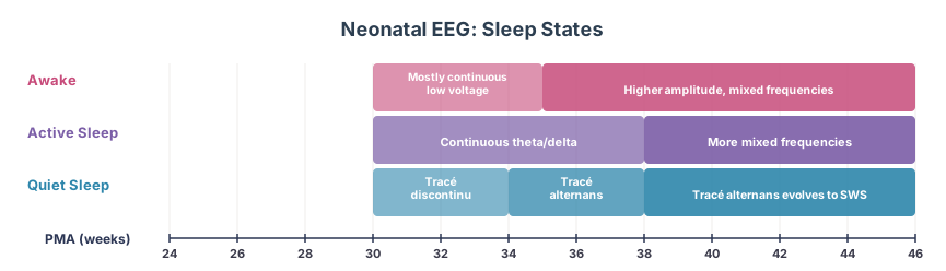
Below is a summary of the changes across the first weeks of life for a neonate. Each will be further discussed in the sections below:
After the first month of life, a baby is no longer a neonate, and becomes an infant. With this there are further expected changes that slowly lead into the tracing of an adult.
Technical Components
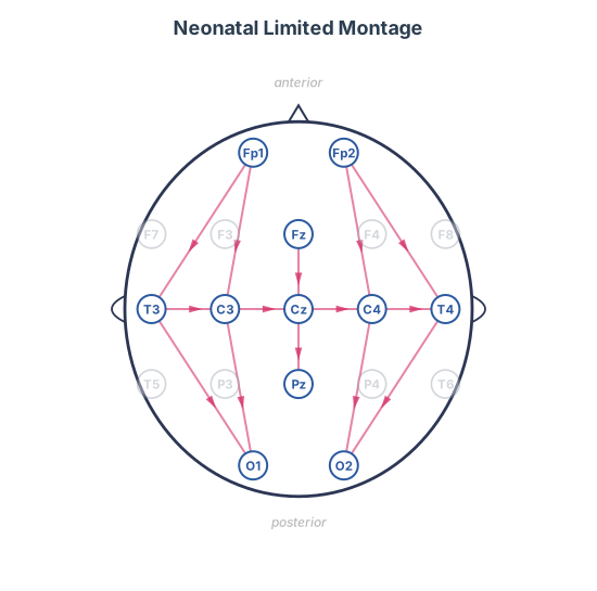
Given the small and sometime alien appearing head size of neonates, a full 10-20 montage puts the electrodes too close together and risks forming salt bridges (discussed in the artifact section) between them. So, the neonatal montage is a pared down version of the 10-20 system that focuses on the central areas, which tend to be more active in neonates, and includes a transverse chain going across from T3 to T4. This version is used until the baby is full term, or at most 46 weeks postmenstrual age (see below for PMA definition).
In the neonate, in addition to the brain and ECG electrodes, there are also extraocular electrodes (electro-oculogram, EOG) placed lateral to the eyes, electromyography electrodes (EMG) on the chin, and leads to assess chest movements for breathing (pneumograph). Neonatal studies are read at a slower page speed of 15mm/sec, with a low frequency filter of 0.01 - 0.5Hz.
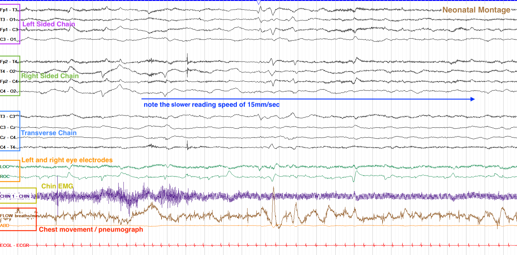
As stated above, neonatal EEGs are starkly different than adults, showing diffuse slowing, discontinuity, asynchrony, and minimal reactivity. Sleep is also different in a neonate, being broken down into quiet and active sleep as opposed to the four stages in adults. These differences, marked as they are, progress into a more classic childhood and then adult EEG, and knowing the timeframe of such changes is critical because what is normal one week for a neonate may be abnormal by the next week.
The timeframe for changes is measured by the postmenstrual age (PMA), rather than chronological age, to take into account prematurity or delayed delivery. The PMA is calculated by adding the gestational age (the time from the mother’s last menstruation, aka the length of the pregnancy) to the chronological age (the time since the baby’s birth). So, a 4 week old baby who was born at 34 weeks would have a PMA of 34 + 4 = 38 weeks.

The postmenstrual age (PMA) is a calculated age that combines the gestational age (essentially, the length of the pregnancy) and the chronological age (time since birth). The PMA should be used when assessing age-based findings on neonatal studies, which change rapidly in the first weeks of life.
Neonatal Background
The background of a neonatal EEG is read across the awake and sleep states, with awake most easily marked by the eyes being open and asleep by the eyes being closed. Differentiable states don’t really emerge, though, until around 29 weeks postmenstrual age; before that, the EEG looks essentially the same all the time. After that, though, states become increasingly apparent as the brain matures rapidly.
Before we delve into neonatal development, let’s review some key terms regarding the background. Continuity refers to continuous waves across the page, without flat periods. Synchrony describes bursts of activity happening at the same time on both sides of the brain (this is different than symmetry, in which the activity is the same in frequency and amplitude on both sides). Reactivity refers to a response or change in the tracing due to external stimuli. Amplitude refers to the height of a given waveform, while frequency (in hertz, Hz) is the number of times a set of waves occurs per second. Morphology describes the shape of a given waveform. In reviewing neonatal studies, you should assess all of these components just as you do with adult studies, so let’s get started.
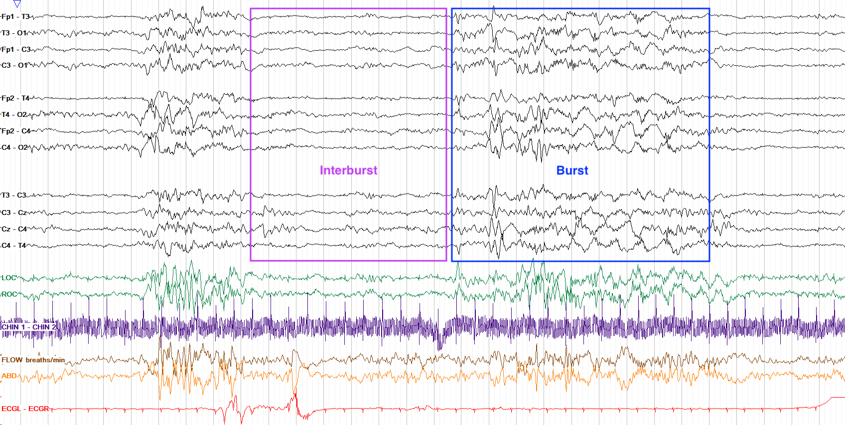
The most striking component of a neonatal background is discontinuity, or periods of essentially flat activity in between bursts of activity. In an adult, such a pattern is highly abnormal, but in neonates as long as the periods of flat activity — termed interburst intervals (IBI) — remain within expected durations (discussed below), it is normal.
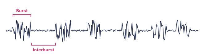
The acceptable IBI starts off quite long and then shortens over time: up to 60 seconds at 24 weeks PMA, 40 seconds at 26 weeks, 20 seconds at 28 weeks, 10-20 seconds from 30-34 weeks, 10 seconds from 34-36 weeks, and 6 seconds from 37-40 weeks (note that these values all fall on the upper limit of normal). After 40 weeks, you can still see some discontinuity in sleep but by 46 weeks at most the tracing should be completely continuous during both sleep and awake states. In the example below, the IBI is about 6 seconds for this 39 week PMA neonate, approximately as expected.
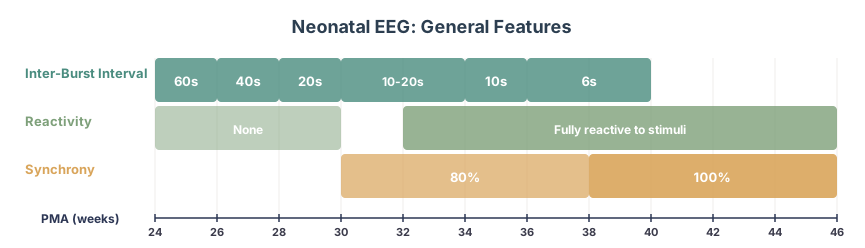
While the IBI decreases across development, synchrony and reactivity increase. Up to 28 weeks PMA a normal tracing may show no reactivity, but starting at 30 weeks you should start to see reactivity to external stimuli; most babies are fully reactive starting between 32 and 40 weeks PMA. Synchrony is achieved somewhat faster, with about 80% of the record becoming synchronous by 30 weeks, and the entire tracing should be synchronous by 38 weeks. This is roughly summarized below:
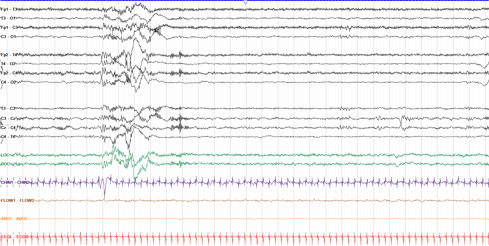
Recall that the max acceptable interburst interval (IBI) decreases across the span of neonatal development, as follows:
24 weeks | 60 seconds
26 weeks | 40 seconds
28 weeks | 20 seconds
30-34 weeks | 10-20 seconds
34-36 weeks | 10 seconds
37-40 weeks | 6 seconds
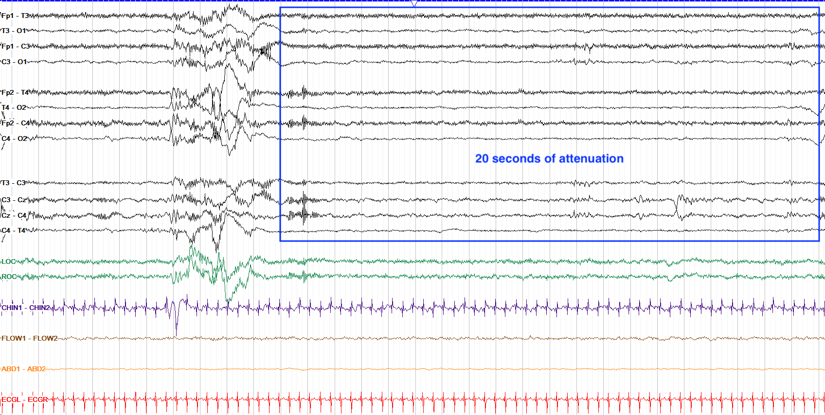
Between 30 and 34 weeks the IBI for most babies drops from 20 to 10 seconds; this baby at 33 weeks PMA would be expected to have closer to a 10 seconds IBI, but we see a 20 second attenuated period across this tracing. So, while technically in the normal range it does show borderline excessive attenuation.
Neonatal States of Consciousness
While neonates before 29 weeks PMA have no discernible states (the tracing is essentially always discontinuous, unreactive and asynchronous), and adults have awake and several phases of sleeping states, neonates after 29 weeks PMA have three states of consciousness: awake, active sleep and quiet sleep.
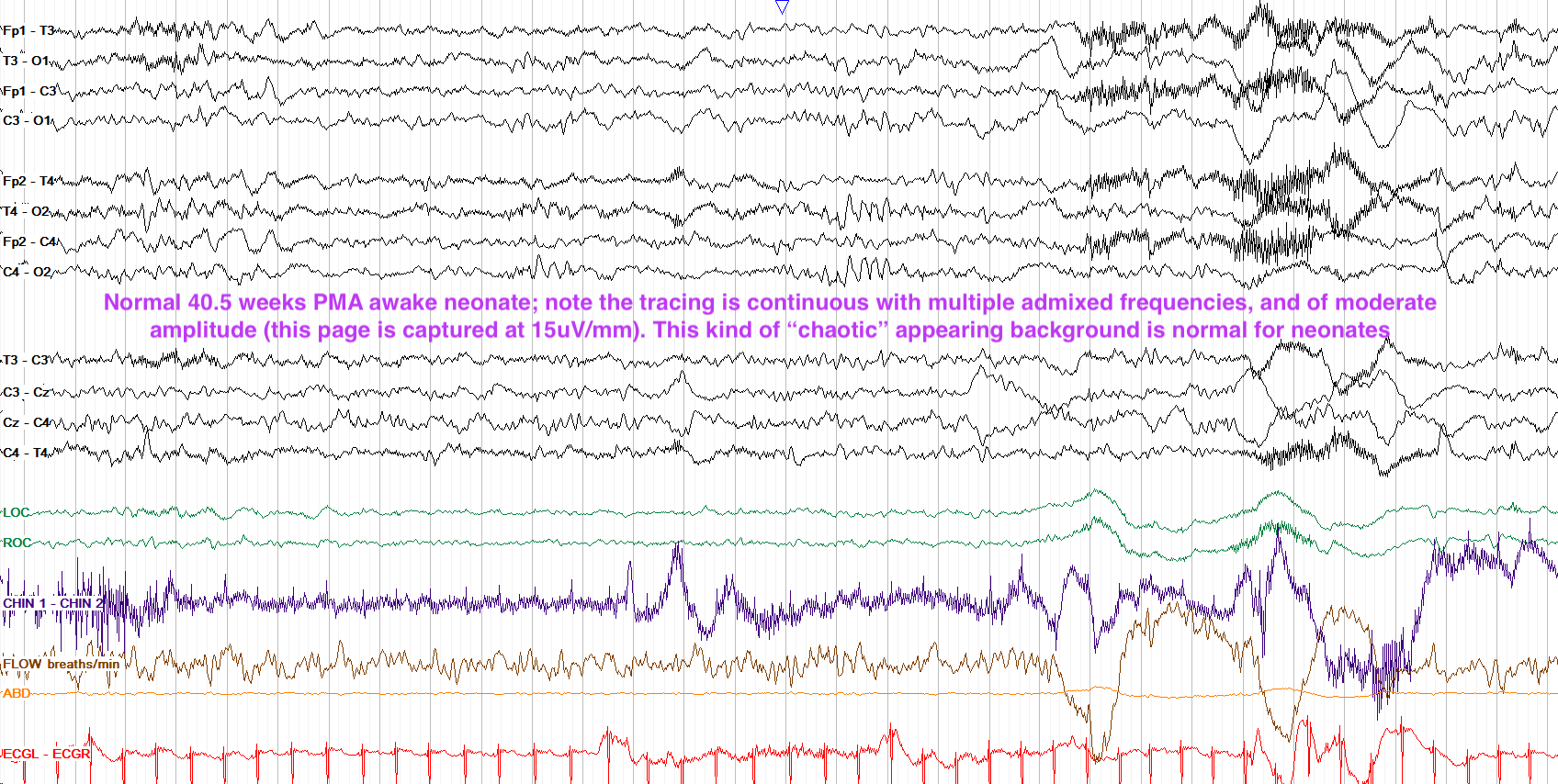
Awake is simply marked by the presence of the eyes being open. The hallmark awake feature from 30-32 weeks PMA is a mostly continuous but rather low voltage background; reactivity and synchrony can vary by age as discussed above. Starting around 35 weeks PMA, the awake background becomes higher amplitude and with a greater mixture of frequencies. Below is a good example of a normal full term neonatal study in the awake state; note the irregular respiratory pattern and movement artifact toward the end of the page that help to confirm the baby is awake.
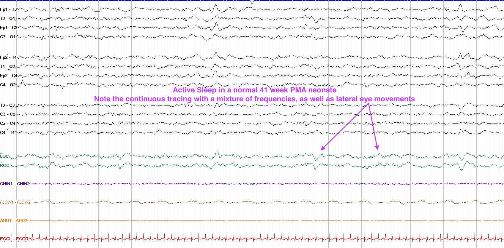
Active sleep is similar to REM in adults, and is marked by the eyes being closed but with other activity such as eye movements, irregular respirations / apneic episodes, and sometimes body movements. From 30 to 36 weeks PMA active sleep is mostly filled by continuous theta to delta activity, and starting at 38 weeks a greater mixture of faster frequencies is also seen. You’ll note that this progression is somewhat similar to the awake state. Below is a good example of active sleep; notice the lateral eye movements seen best in the LOC and ROC leads, the continuity of the tracing with mixed frequencies, and the lack of movement artifact.
Quiet sleep is characterized by the eyes being closed with minimal eye movements and regular respirations. Generally, neonates move from awake into active sleep, then into quiet sleep (they spend about 50/50 in each type of sleep).
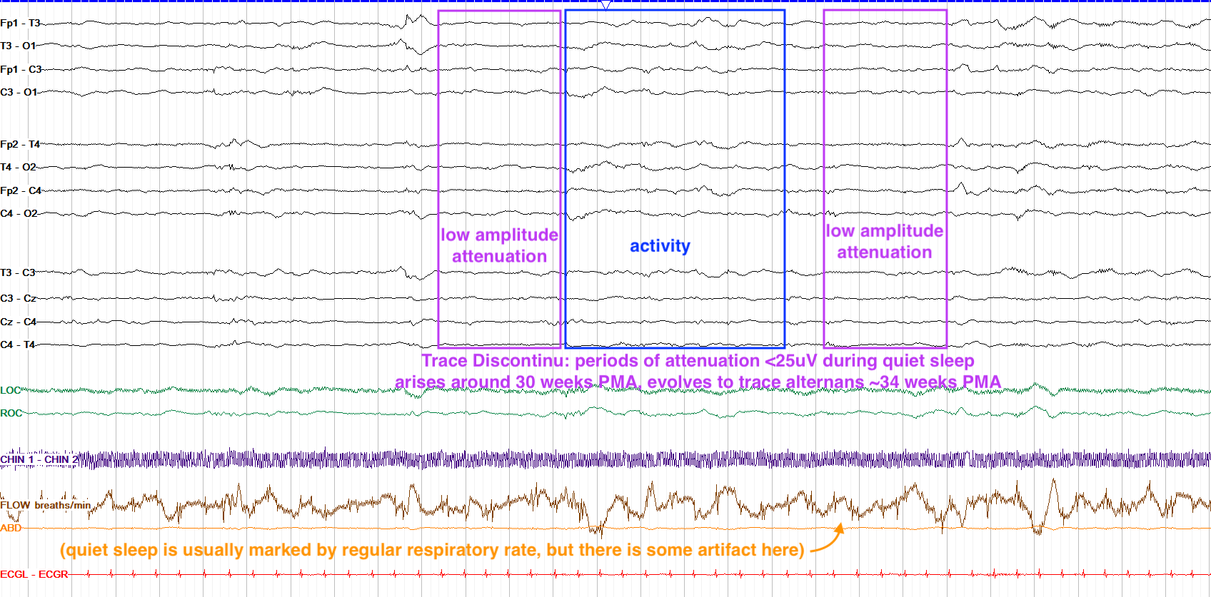
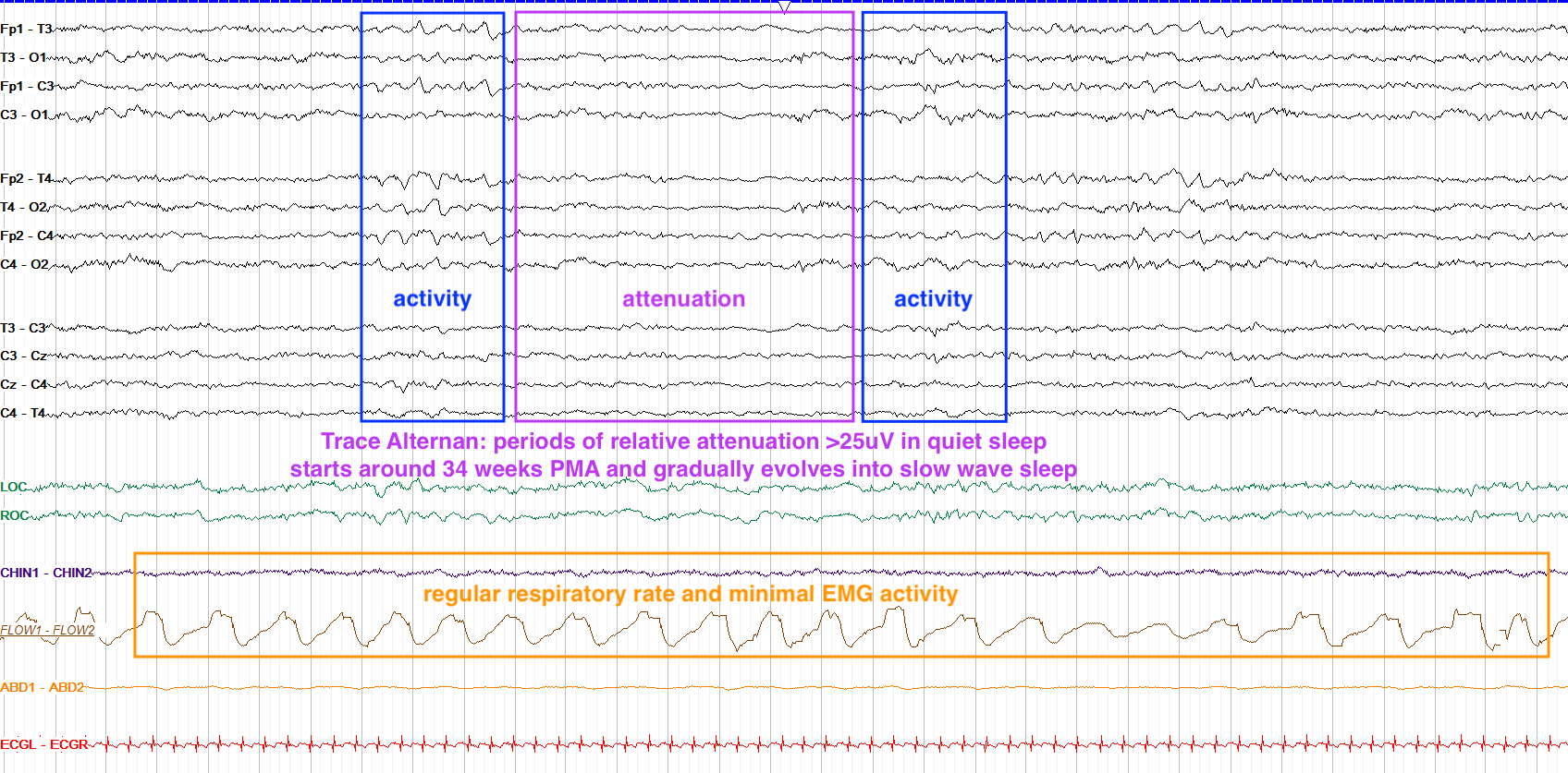
From 30-34 weeks PMA, quiet sleep is predominated by a tracé discontinu pattern, marked by bursts with interburst intervals, lasting 15 seconds or less, with the interburst intervals having amplitude less than 25 microvolts. Starting at 34 weeks PMA, tracé discontinu evolves into tracé alternans, in which interburst intervals are higher amplitude — 25 microvolts or above — and the IBI continues to shorten.
From 38 weeks PMA onward tracé alternans continues to evolve into slow wave sleep. To summarize, discontinu and alternans differ primarily in their interburst interval amplitude (discontinu is <25 and alternans is >25 microvolts), and alternans is a more mature pattern; the examples below (both captured at the same sensitivity) may help provide a better sense of the difference.
Between awake and quiet sleep, there are often periods of transition that can be unclear, but as development continues the states should become more clearly defined; such periods of indeterminate sleep may be seen more often in neonates with brain injury, such as hypoxic ischemic encephalopathy (HIE).
Being able to recognize the state of a baby is equally important as it is for adults, albeit often less clear cut. Take a minute to review the expected timeline for state changes, remembering that prior to 28-30 weeks there really aren’t any different states:
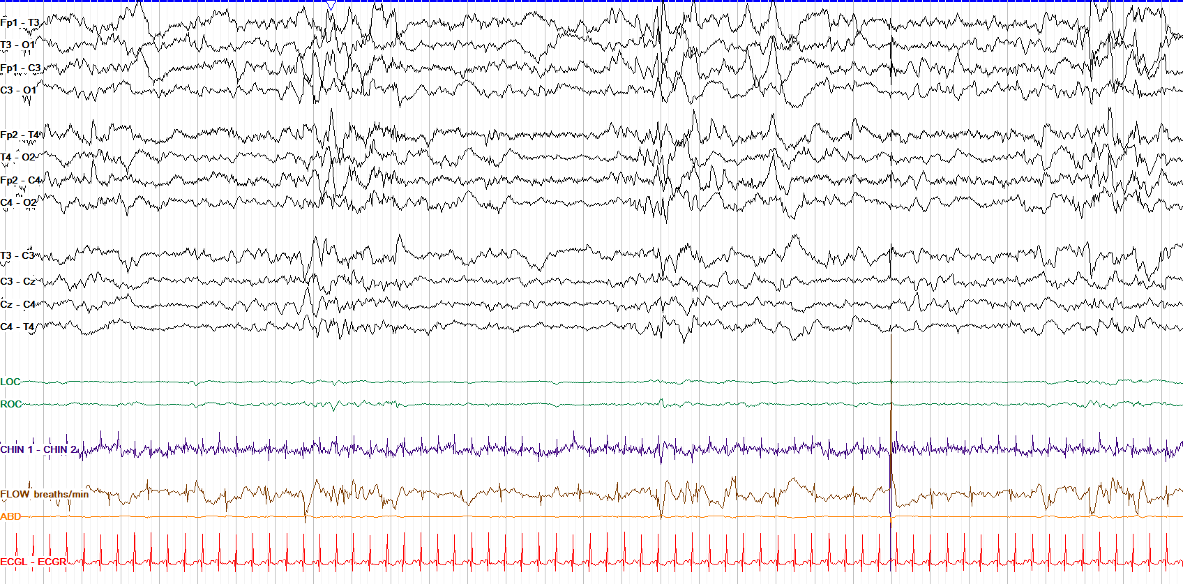
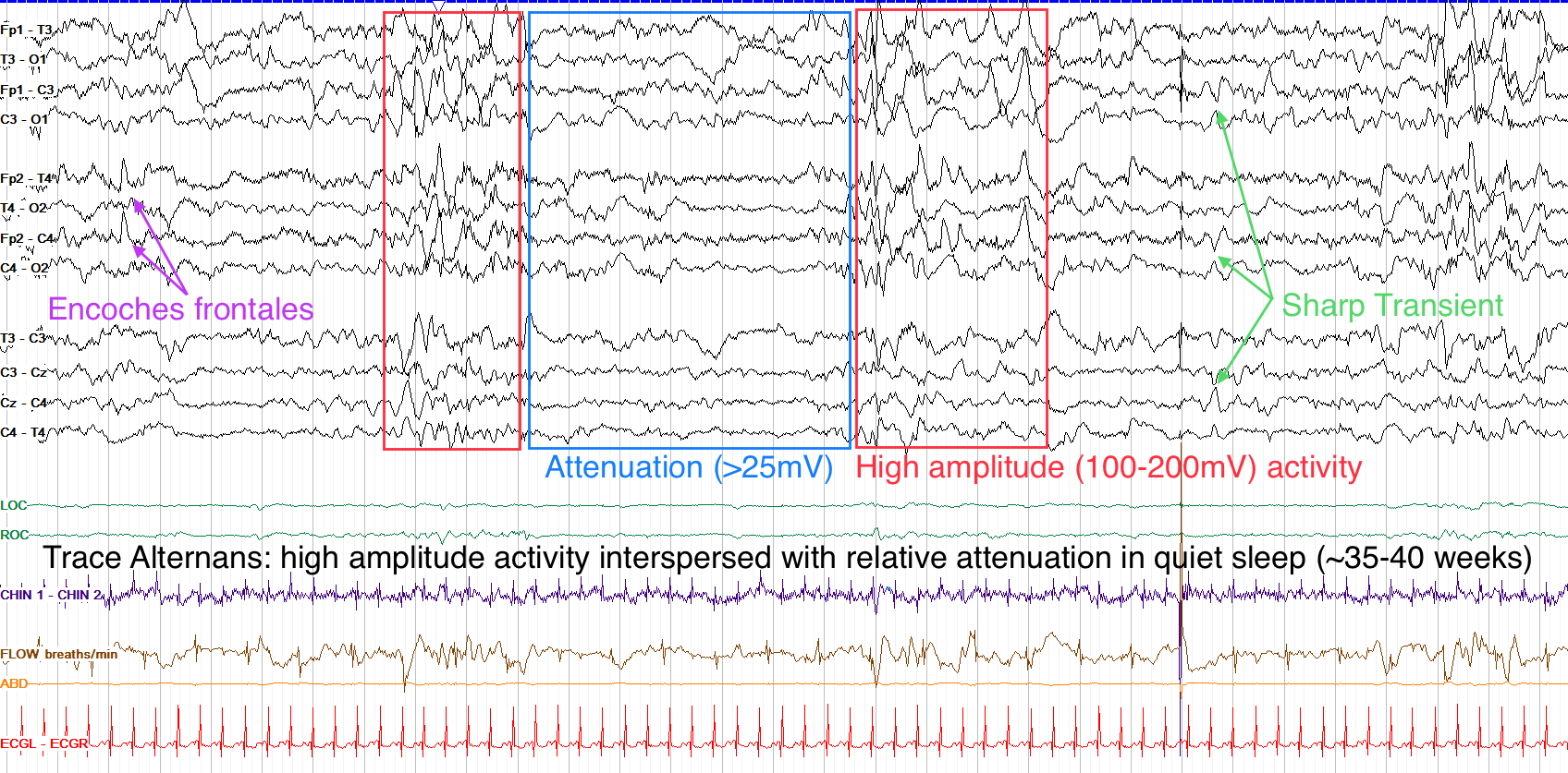
Recall that by 40 weeks the background for an awake neonate should be largely continuous, but periods of discontinuity can remain during quiet sleep. On this tracing we see periods of attenuation between higher amplitude activity, and while we don't have a measurement of the actual voltage, the attenuated periods are not too flat, likely most consistent with a tracé alternans pattern. As such, this baby is in quiet sleep. Note also the lack of eye movements seen in the LOC and ROC leads, and the presence of mostly just ECG artifact in the EMG chin lead; both of these also support quiet sleep. There are a few sharp transients here too, which are discussed more below.
Neonatal Transients
Transients (also termed graphoelements) describe particular waveforms that are seen only in neonatal studies, many of which could be mistaken for abnormalities to the inexperienced eye. Of course, with your ever-increasing EEG expertise, you won’t have that problem. As with all elements of the neonatal study, normal transients vary by age.
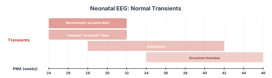
The earliest transients seen are temporal sawtooth theta and monomorphic occipital delta. Both start around 24 weeks PMA and last until around 30-32 weeks PMA. They both also have very descriptive names; with temporal sawtooth theta, look for sharply contoured, moderate to high amplitude bursts of theta activity in the bitemporal regions, while monomorphic occipital delta appears as its name suggests. Both of these patterns are really only seen in very premature babies, as they fade away before delivery for most neonates.
Delta brush, on the other hand, is a finding you may see in only mildly premature newborns, and describes 8-20 Hz fast activity overriding delta waves, like the bristles on a brush. It arises around 28 weeks rather diffusely, should become mostly posterior by 36 weeks, and usually goes away completely by 40 or, at most, 42 weeks. While delta brush is a normal part of neonatal EEG, you may also find it present in the abnormal tracings of adult patients with NMDA receptor encephalitis.
Starting around 34 weeks PMA, frontal sharp transients, or encoches frontales, arise. These are bifrontal and synchronous frontal discharges that are normal from 34 weeks to up to 46 weeks. They really should be synchronous, as lack of synchronicity for them suggests it to instead be an abnormal epileptiform discharge.
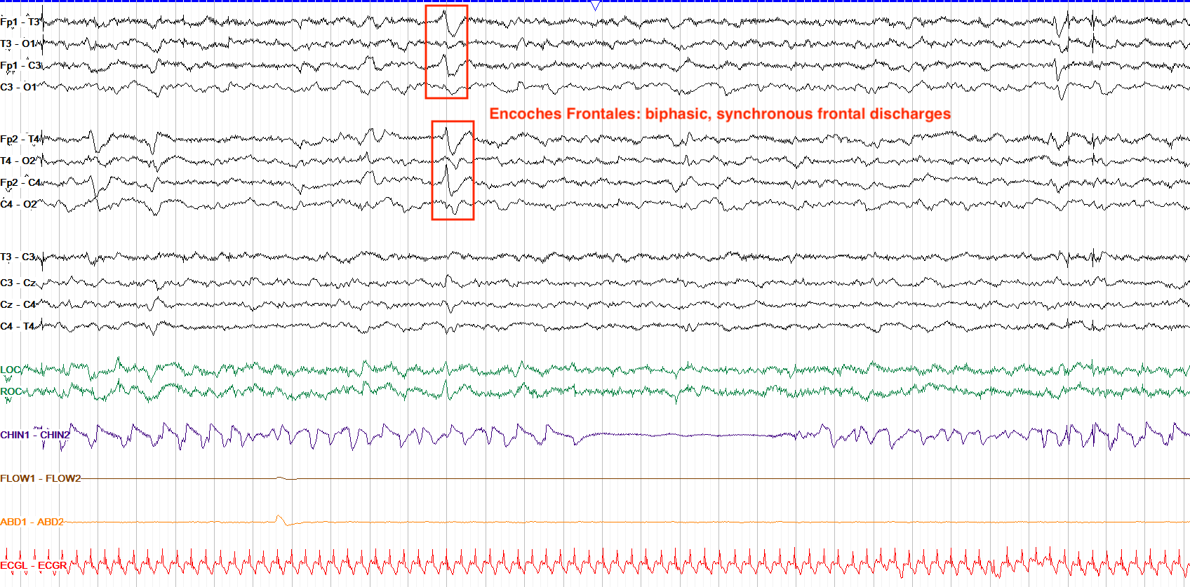
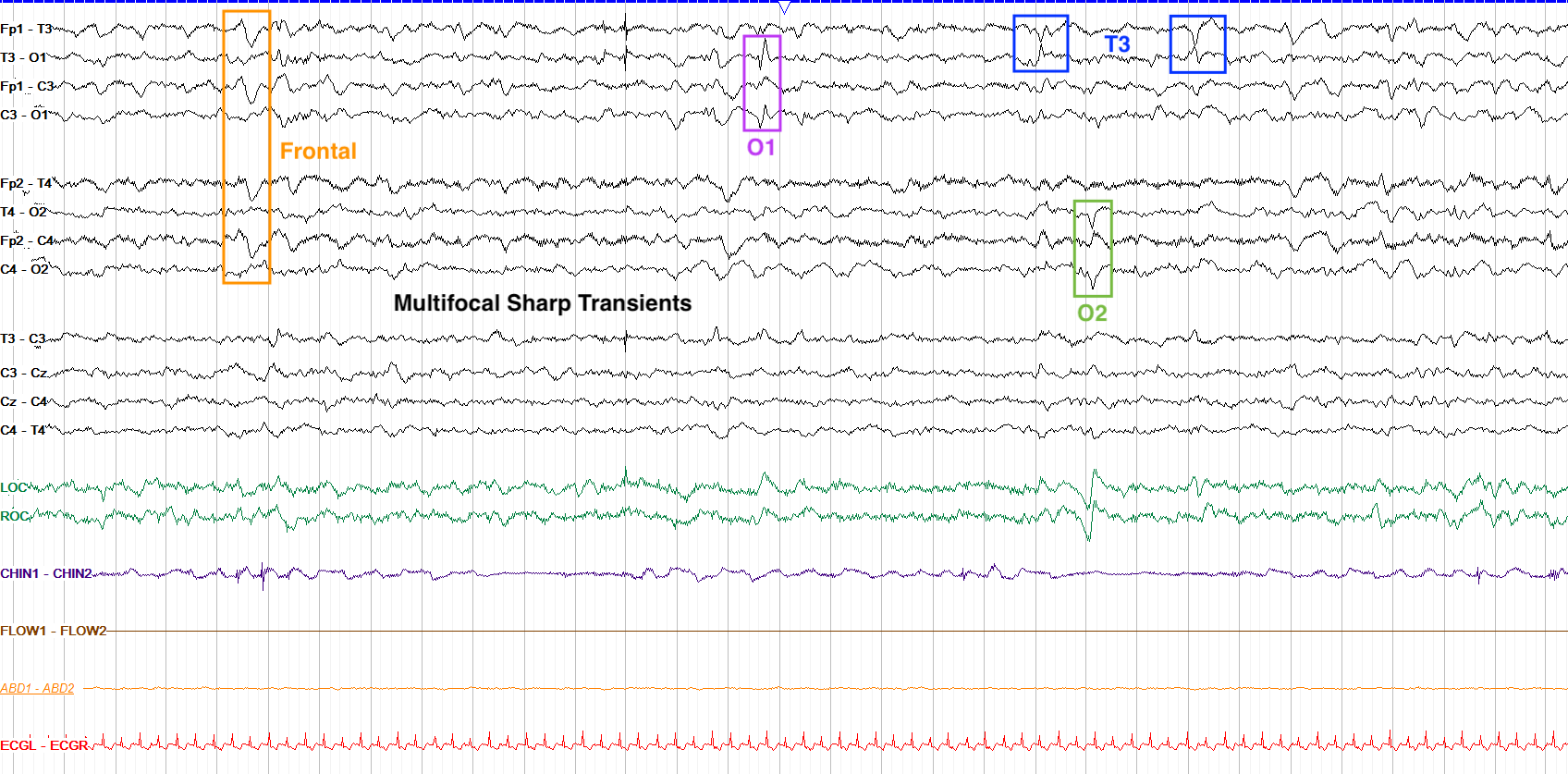
Aside from frontal sharp transients, centrotemporal sharp transients are also normally seen, while more widespread and multifocal sharp transients are usually normal within the first month of life, but should fade away by 49 weeks PMA. On the example below from a healthy 39 week PMA baby, note the numerous sharp transients in various locations; as long as a particular location doesn’t become excessive in sharps, this is most likely normal.
Below is a summary of the more commonly seen neonatal transients and their expected timelines:
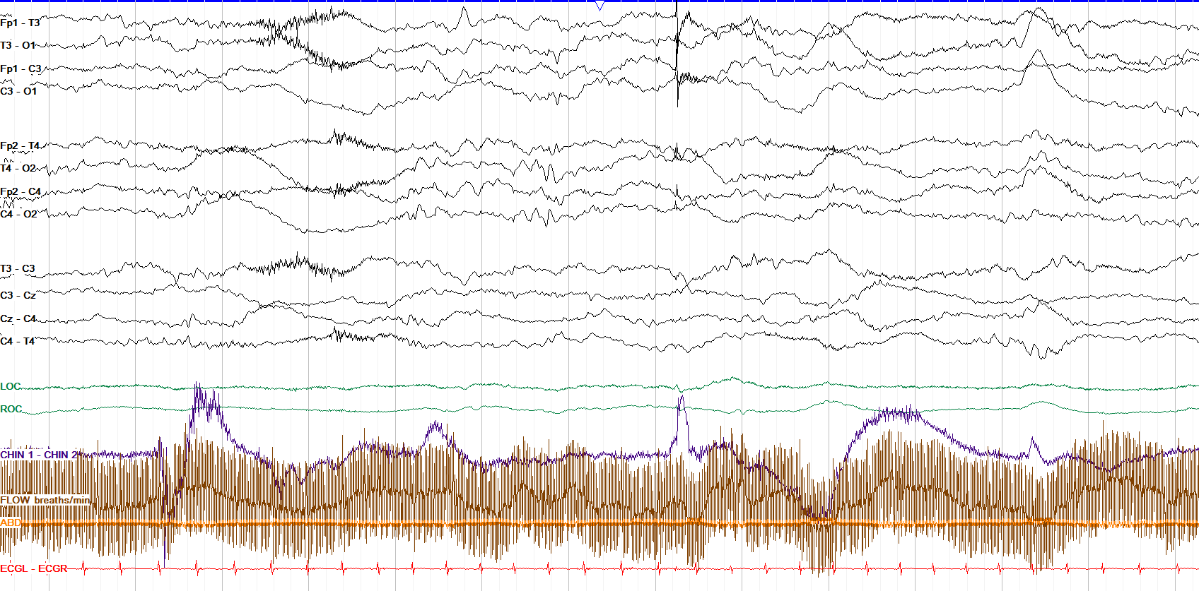
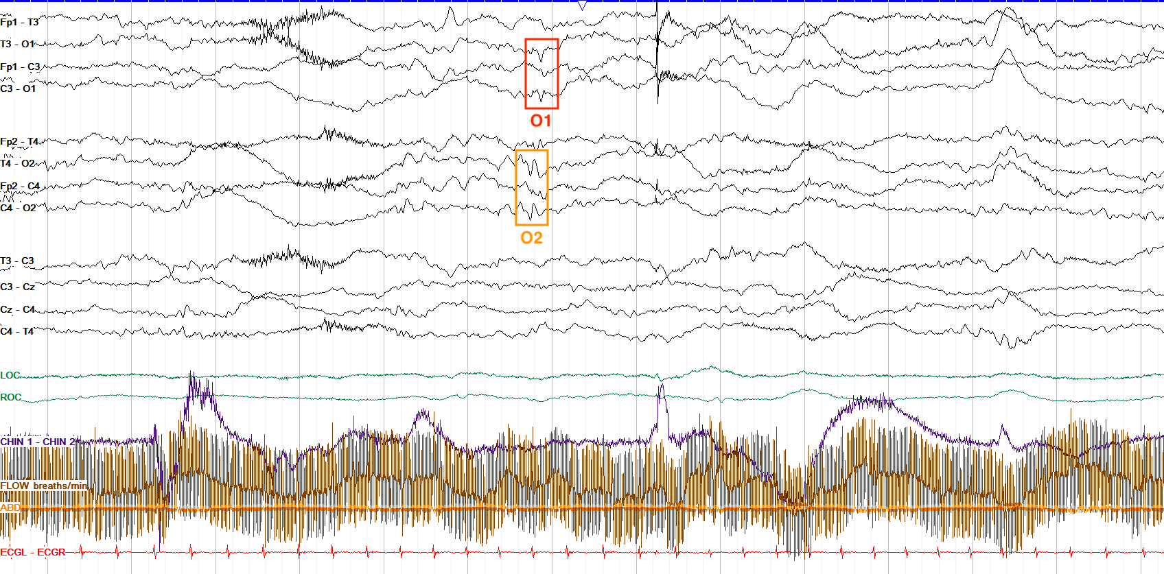
This tracing has a lot of movement and other artifact on it, but you can see right near the middle a clear spike at O2 and, about 1/5 of a second later, a smaller spike at O1. Both of these are consistent with sharp transients although the O2 discharge has a slightly spike wave morphology. It is possible that with a bit more brain maturation, these would become a synchronous, symmetric bioccipital discharge. With such a finding you’d want to continue looking for more to ensure that these don’t become very frequent, persistent, or strongly lateralized, which would be more concerning for epileptiform activity. Note that this tracing has an improper reading speed for neonatal studies — they should be read at 15mm/sec, and this page is at the adult speed of 30mm/sec.
Neonatal Epileptiform Abnormalities
Epileptiform activity in neonates follows the same rules as it does for adults — it tends to be negative in polarity, the classic spike and slow wave morphology is often present, and it should disturb the background with a field. The difficulty with differentiating neonatal epileptiform activity comes mostly from the fact that it exists among the many other normal neonatal transients we discussed above, and you need to pick out one group from the other.
As mentioned already, the location of sharp waves in neonates can be helpful in determining if they are epileptiform or not: frontal and centrotemporal sharps are probably benign, while midline and occipital sharps are probably epileptiform. Rare multifocal spikes (a few per hour) are often normal. As with any rule of thumb, there are exceptions; for example, an excess of frontal or other localized sharps, persistently focal sharps, or asymmetric discharges in any location should raise alarm for true epileptiform activity rather than just normal transients. Furthermore, spikes in the awake or active sleep state after about 42 weeks PMA are more likely to be abnormal.
Similar to adults, persistent rhythmic activity can also be concerning as epileptiform activity in neonates. Normally, you can see centrotemporal bursts of rhythmic activity, or randomly localized rhythmic activity. If you start to see a particular region with consistent rhythmicity, though, be suspicious of abnormality.
Aside from interictal activity, neonatal seizures are particularly important to see on EEG because many neonatal seizures are clinically silent—EEG is the only way you’ll know they’re happening. Fortunately, seizures in neonates follow the same rules as those for adults: they must evolve.
Generally, focal or multifocal seizures are more common than generalized seizures in neonates. They can, however, be easy to pass by on screening because they can be relatively low amplitude with longer sharps; their rhythmicity or periodicity, though, may be more apparent. For neonates, particularly, seizures can come with desaturation events or other very subtle clinical findings. Below is an example of a right hemispheric onset neonatal seizure:
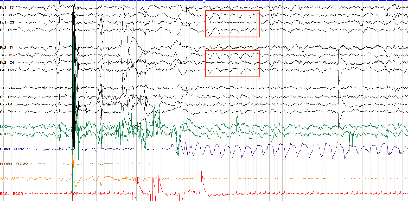
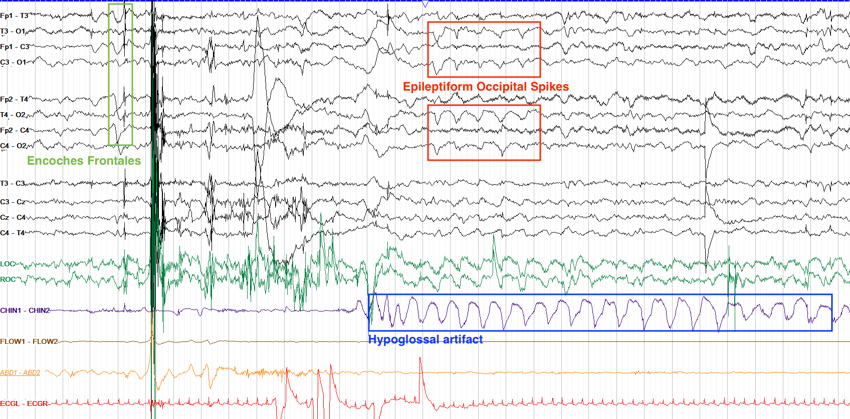
While neonatal transients can sometimes occur in the occipital regions, the marked waveforms are a rhythmic run of spike wave discharges lasting four seconds, which is not consistent with normal transients. Note that we do see some normal transients here via encoches frontales — synchronous bifrontal discharges — in the first few seconds of the page. There is also some hypoglossal artifact picked up on the EMG lead, which could cause rhythmic artifact on the EEG but in this case the hypoglossal activity 1) doesn’t match up timewise to the occipital discharges and 2) the occipital discharges have a spike wave morphology that makes them less likely to be just artifact.
- The neonatal EEG rapidly evolves, reflecting the maturation of the brain, and uses a limited montage with a focus on the midline and vertex regions.
- Babies become reactive to stimulation from ~30 weeks onward, and reach full synchrony by ~38 weeks PMA.
- Discontinuity is common in very early neonates, but by 40 weeks PMA discontinuity should be seen only in quiet sleep with very short (<6 seconds) periods of attenuation.
- Neonates have three states of consciousness: awake, active sleep and quiet sleep.
- Awake and active sleep look similar, with theta and delta frequencies that become continuous by 40 weeks PMA; the main difference is that the eyes are closed in active sleep and open when awake.
- Quiet sleep has two phases: from 30 to 34 weeks PMA it shows tracé discontinu (attenuated periods <25μV) and from 34 to 38 weeks PMA it shows tracé alternans (attenuated periods >25μV) that evolves into slow wave sleep from 38 weeks PMA onward.
- Temporal sawtooth theta and monomorphic occipital delta are seen in very premature infants and subside by around 32 weeks PMA.
- Encoches frontales (to ~46 weeks) and multifocal sharp transients (to ~49 weeks) persist into the term period, while delta brush resolves by ~40-42 weeks PMA.
- Discharges that are extremely frequent, persistently in one area, or become rhythmic are concerning for epileptiform activity rather than just sharp transients.
- In neonates, occipital and midline sharps are more likely to be abnormal than frontal or centrotemporal sharps.
- Neonatal seizures must evolve just like adult seizures do, and tend to be focal or multifocal rather than generalized.