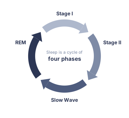
The asleep EEG goes through four phases: stage I, stage II, slow wave, and rapid eye movement (REM), each with characteristic EEG findings.
Stage I Sleep
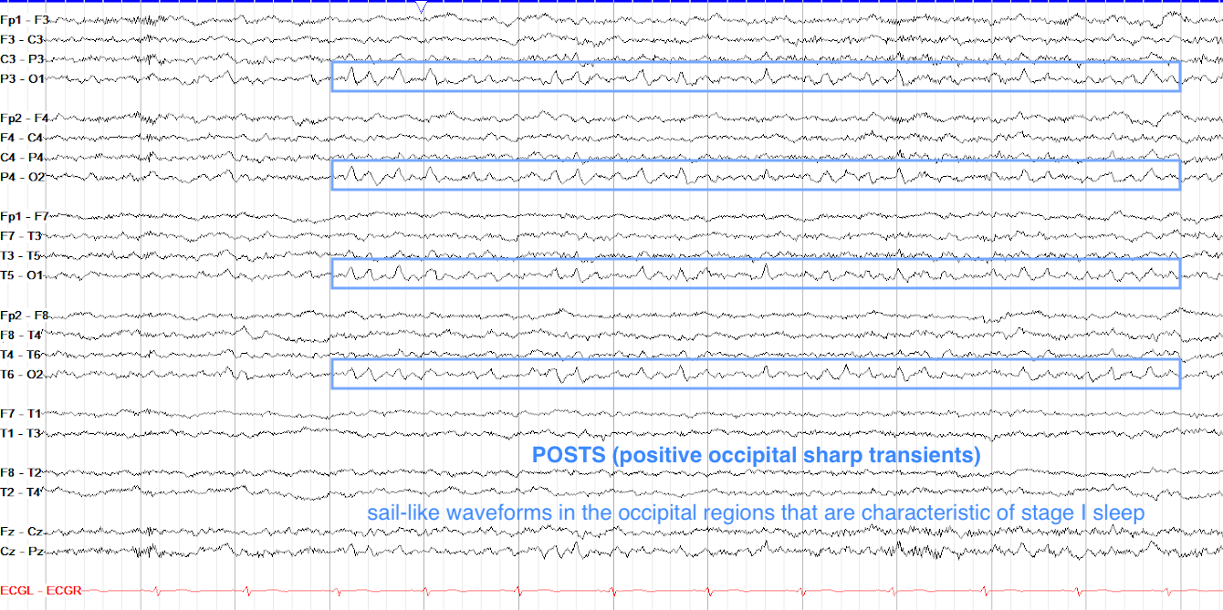
Stage I sleep (N1 in current AASM staging) is somewhere along the spectrum from drowsiness to being fully asleep, and is marked most easily by the appearance of drowsiness (emergence of theta, decreased eye blinks, slow roving eye movements as discussed in the awake section), then a gradual loss of the PDR with coinciding diffuse attenuation of the tracing, and finally the presence of positive occipital sharp transients of sleep (POSTS) and vertex waves.
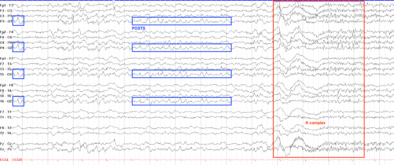
POSTS are a very descriptive waveform, as they are positive deflections seen in the occipital leads that look like sharps, but are not epileptiform, as seen below. Classically, they have a "sail-like" appearance, and can come in singles or runs. They arise in stage I sleep, but can persist throughout the later stages of sleep. POSTS have a cousin in the awake state called lambda waves, which are discussed in the artifact section.
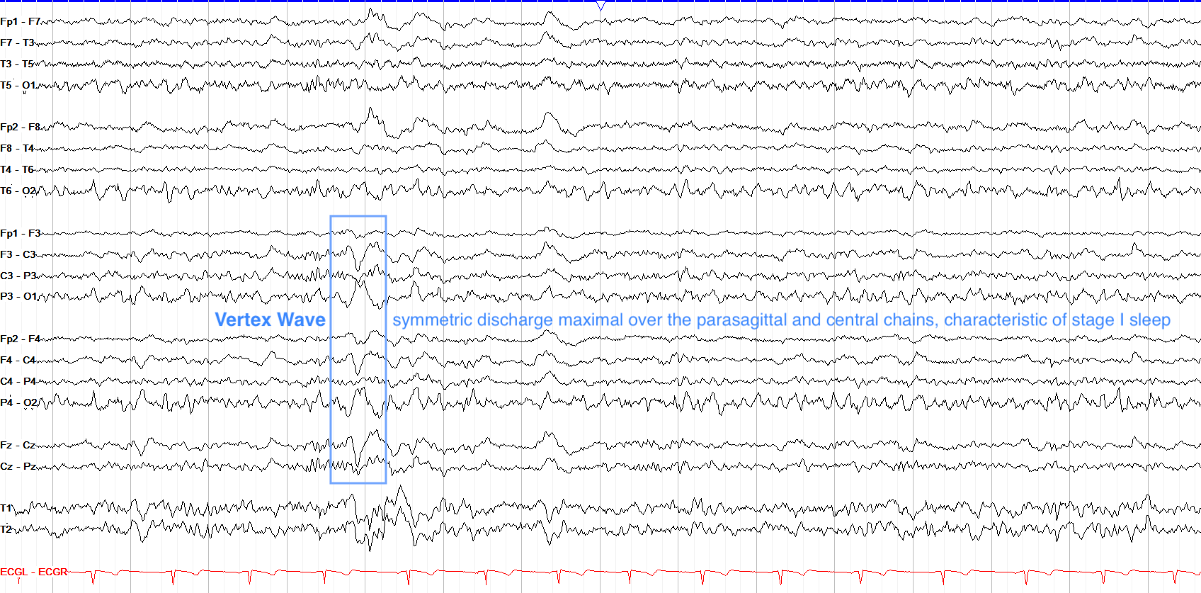
Also found in stage I sleep are vertex waves, which are bilateral phase reversing discharges over the central regions. They can come alone or in runs of varying amplitude and morphology, but even when they appear very sharp or high amplitude they are not epileptiform. In children, vertex waves can be quite prominent and sharply contoured but still remain normal, as discussed in the pediatric section.
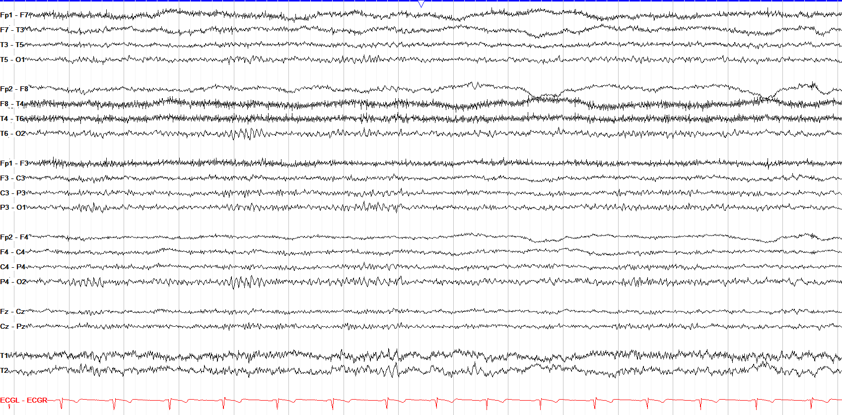
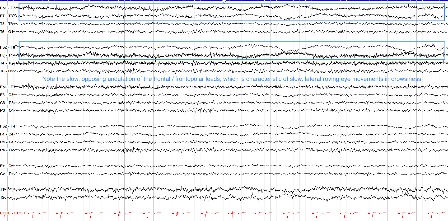
This patient is awake but very drowsy. Recall that drowsiness is marked by diffuse attenuation and possibly mild slowing of the background, but you can still see a clear PDR in the posterior leads here. In the frontal and frontopolar regions, opposing slow undulations are seen in polarity, indicative of lateral roving eye movements. This occurs because the cornea is positively charged, and thus when you look to the right, the right eye's cornea gets closer to F8 and it sees a positive charge; at the same time, the left cornea moves away from F7 and thus it sees a negative charge. So, lateral eye movements lead to a frontal positive charge on the side to which you're looking, and a negative charge on the opposite side.
Stage II Sleep
Stage II sleep (N2) is characterized by the arrival of sleep spindles and K complexes, although both vertex waves and POSTS persist into stage II from stage I sleep.
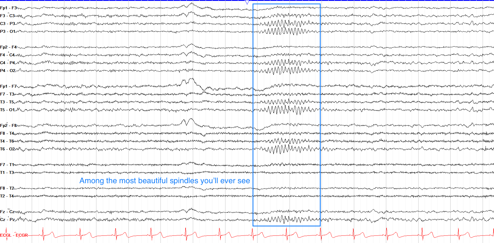
Sleep spindles are symmetric bursts of 11-16 Hz activity arising from the reticular nucleus of the thalamus; they are so named because they can look like spindles of thread, as seen below. The absence of spindles, or an asymmetry of their formation or frequency, can be abnormal.
K complexes are higher amplitude, symmetric waveforms with an initial negative followed by a slow positive phase, often but not always followed by a sleep spindle. They are named K complexes because in early sleep studies researchers noted that knocks or other noises while a patient was asleep brought them on.
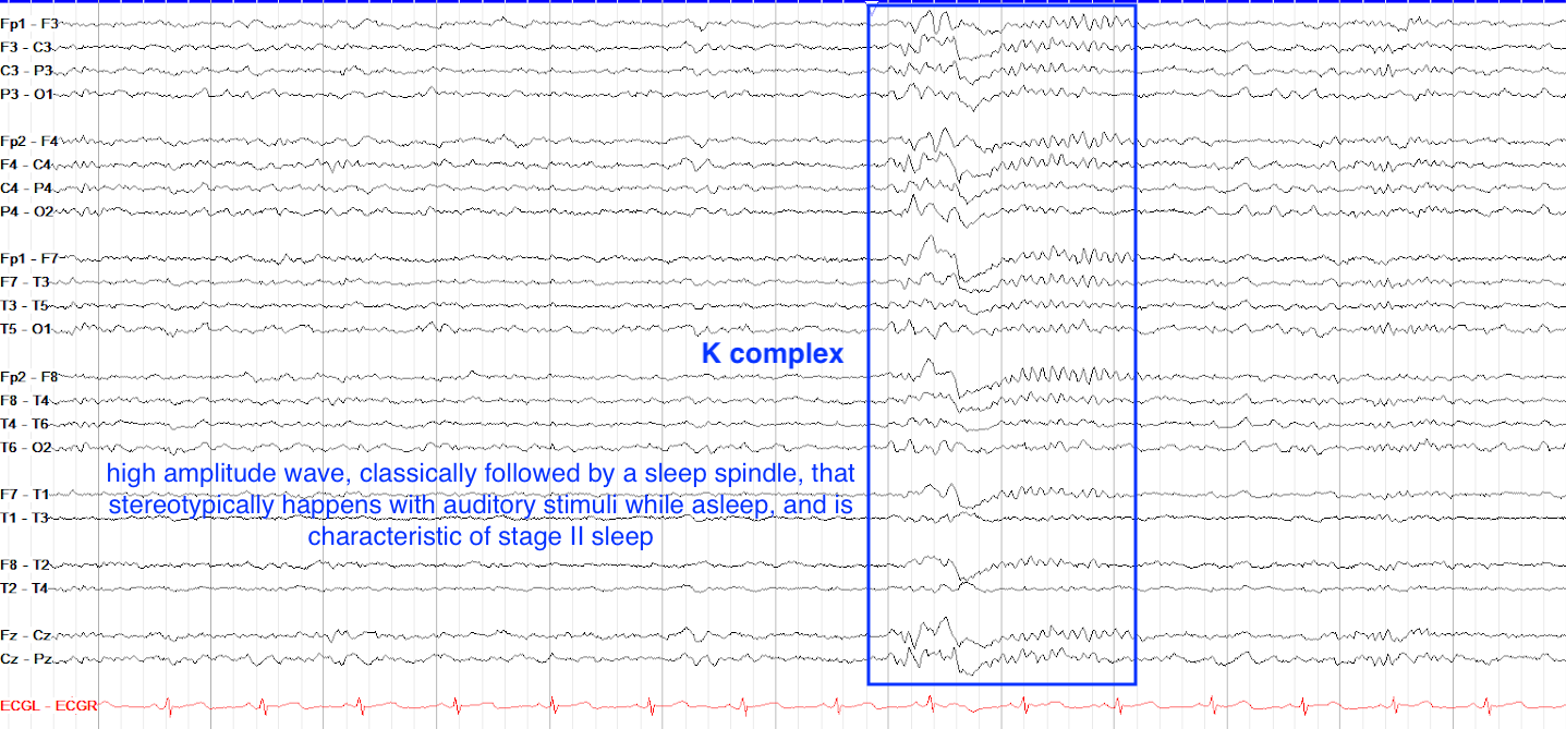
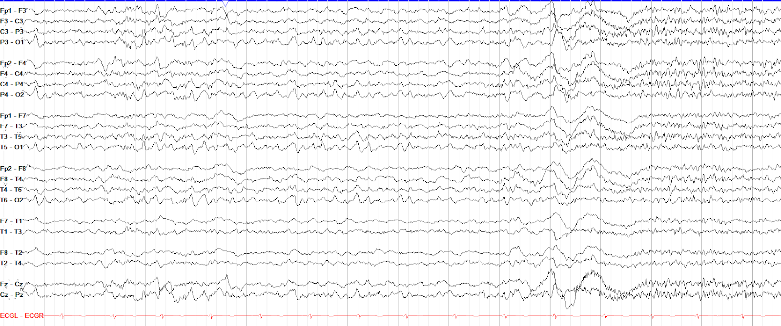
Stage II sleep is characterized by all of the architecture from stage I -- including vertex waves and POSTS -- along with the arrival of sleep spindles and K complexes. Note the prevalence of POSTS on the first half of this page, then the high amplitude, diffuse and biphasic K complex followed quickly by an 11-16 Hz diffuse sleep spindle. This is an excellent example of a normal sleep tracing. There may also be a poorly formed vertex wave seen in the parasagittal leads in the fourth second of the page.
Slow Wave Sleep
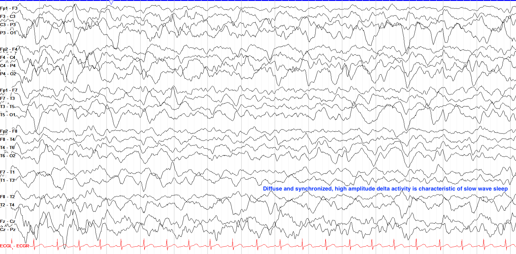
Slow wave sleep (N3, and previously split into stages III and IV) is marked by high amplitude (>75 µV), synchronized delta activity, usually 0.5-2 Hz in frequency. All of the architecture from earlier sleep phases (vertex waves, POSTS, spindles and K complexes) can be present in slow wave sleep, but usually become somewhat less organized, and often disappear altogether. Slow wave sleep can appear to be extremely high voltage, particularly in younger patients. Some medications, such as benzodiazepines and barbiturates, reduce the proportion of slow wave sleep.
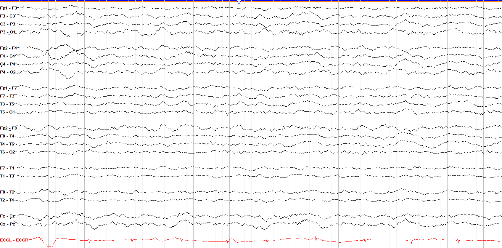
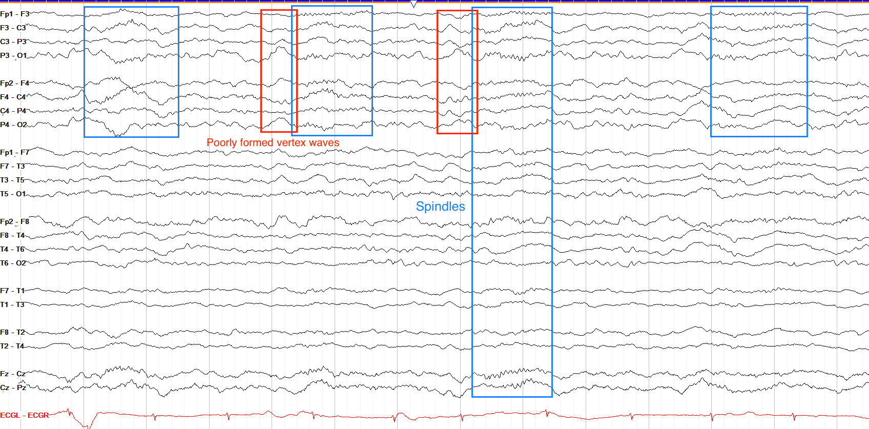
The presence of somewhat blunted but phase-reversing vertex waves broadly over the central regions and diffuse spindles (albeit subtle and lower amplitude compared to prior examples) suggests that we're in at least stage II sleep here, but there is a bit more delta than we see in earlier stages of sleep and the morphologies of the stage I and II architecture are not as crisp as past examples. Thus this is most consistent with stage II sleep progressing into early slow wave sleep.
Rapid Eye Movement Sleep
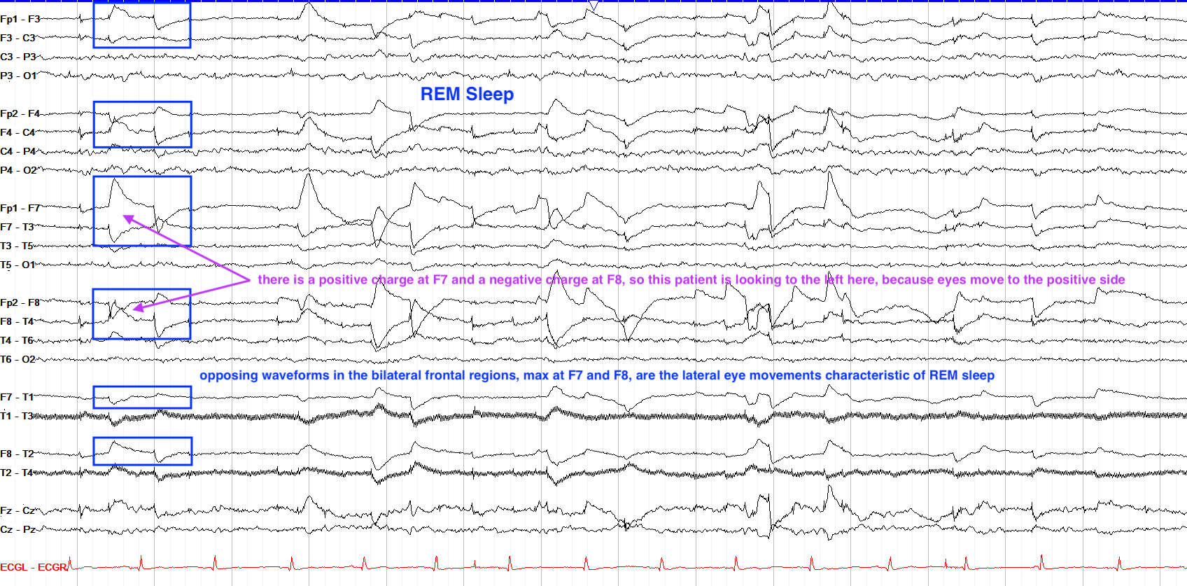
Rapid eye movement, or REM sleep, increases in proportion across the span of the night. It is marked by a diffuse attenuation of amplitudes, with a range of frequencies amongst the background that can look similar to an awake state. It derives its name from rapid eye movements that arise and are seen on EEG as very sharply contoured, opposing left and right frontal waveforms with a faster upslope than downslope.
These waves emerge because the cornea is positively charged, so when a patient looks to their right, the right eye cornea moves closer to the F8 electrode, leading to a positive charge seen at F8; at the same time on the left side, the left eye's cornea moves away from the F7 electrode, leading to a negative charge seen at F7. Thus, opposing frontal waveforms are seen: a positive wave on the side to which you're looking, and a negative wave on the opposite side. A nice mnemonic for this: look on the bright (positive) side.
In REM sleep one can also see some mild cardiac irregularity and breathing fluctuations, and muscle activity should be near-absent throughout this stage.
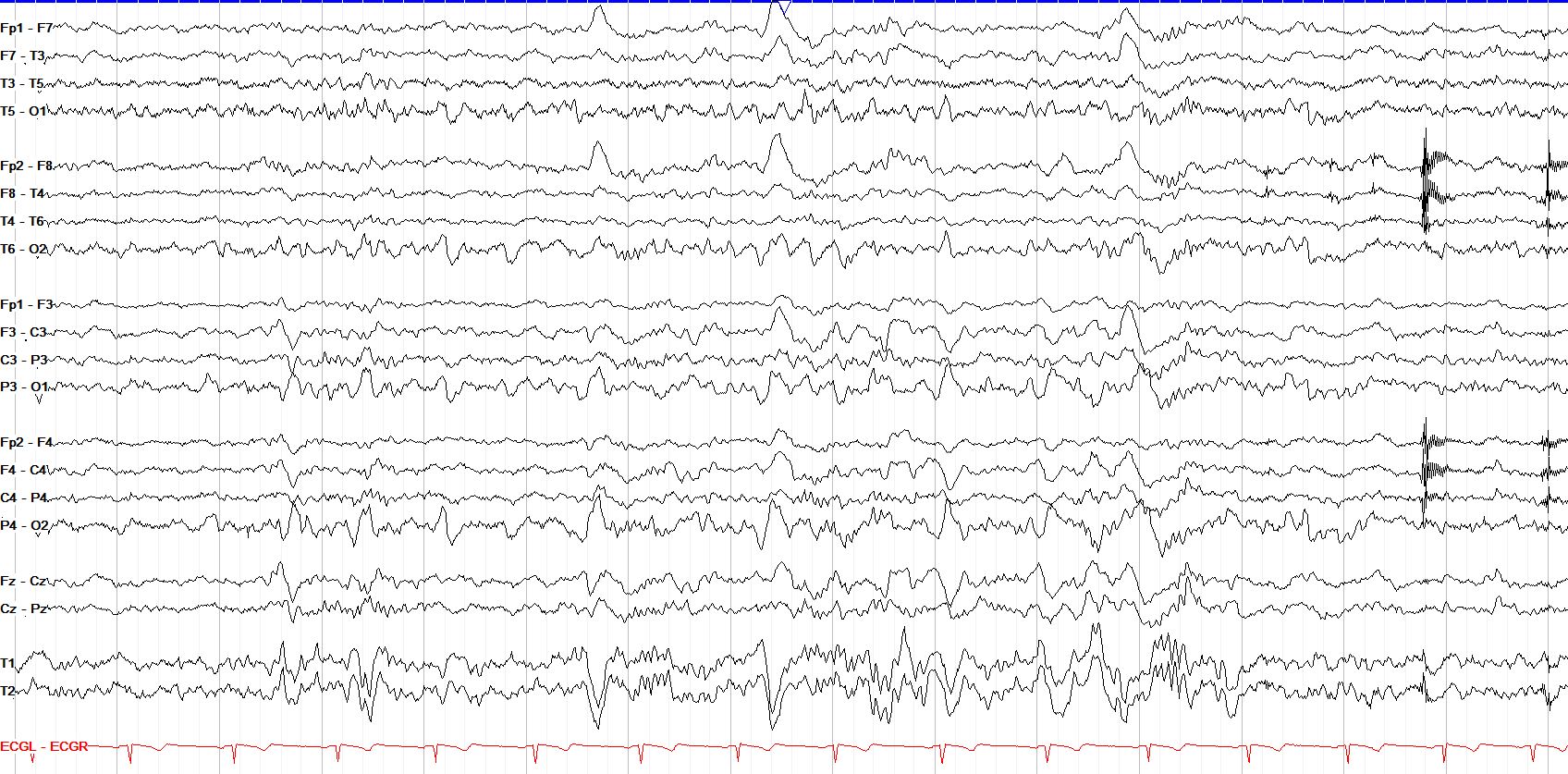
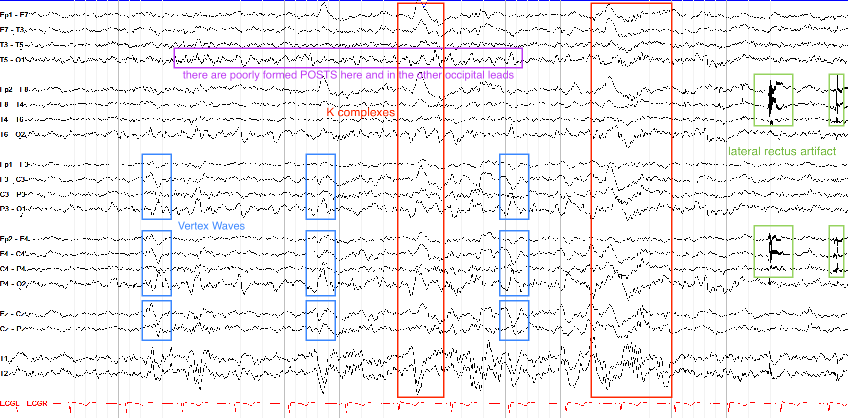
This page shows several clear vertex waves, marked by high amplitude and symmetric discharges in the parasagittal regions and centrally, with a few less well formed ones in between the ones marked in blue boxes. There are also poorly formed POSTS in the occipital regions bilaterally. This tracing appears to be in the initial transition from stage I to stage II sleep, as some of the higher amplitude background activity in the middle and towards the end of the page are K complexes. Of note, the frontal bursts of very high frequency activity marked in green are lateral rectus muscle artifact (which are more classically seen in REM sleep); these are discussed in the artifact section.
- Sleep is a series of four phases: stage I, stage II, slow wave, rapid eye movement (REM).
- Stage I sleep is marked by vertex waves and positive occipital sharp transients of sleep (POSTS).
- Stage II sleep is marked by sleep spindles and K complexes.
- Slow wave sleep is marked by diffuse, synchronized, high-amplitude (>75 µV) delta activity at 0.5-2 Hz.
- REM sleep has diffuse attenuation of activity with lateral eye movements in the frontal leads.