Don’t Overcall Normal Variants
Aside from the usual background findings and artifacts, EEGs will often have normal cerebral patterns that, to an untrained eye, may appear to be epileptiform or otherwise pathological; this can lead to a normal tracing being incorrectly read as abnormal, and subsequently to unnecessary tests or treatments. Learning to accurately recognize and differentiate these normal variants is thus critical to balanced EEG interpretation. Technically these findings could be included in the normal awake and asleep sections, but given that they aren’t always seen they are not requisites for describing the background.
Mu Rhythm
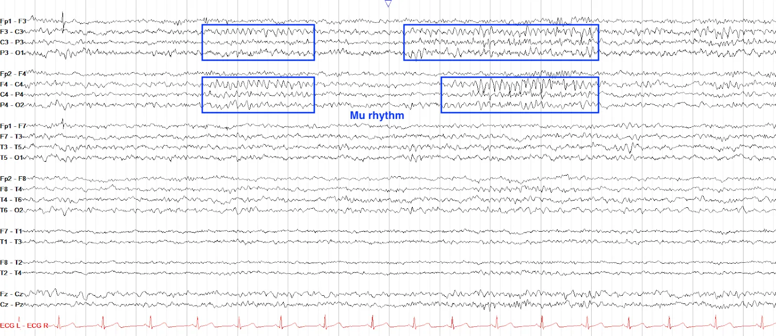
The Mu rhythm is the idling activity of the sensorimotor cortex, similar to how the PDR is the idling rhythm of the occipital regions. Mu appears as arch-like alpha activity (usually 7-11 Hz) over the centroparietal (central) regions along the parasagittal chains; it can be bilateral but is often predominant on one side or the other. Particularly in cases of breach rhythm, it can appear rather high amplitude and sharp, but should not be mistaken for epileptiform activity.
Mu is suppressed with thinking about or actually initiating motor activity, so is often not present (i.e. if you think of moving the right arm, the left Mu will recede).
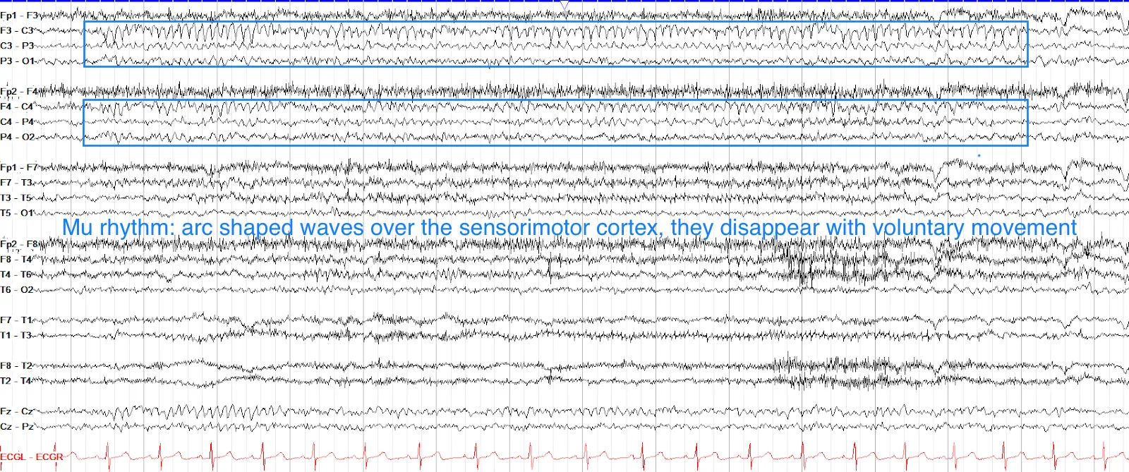
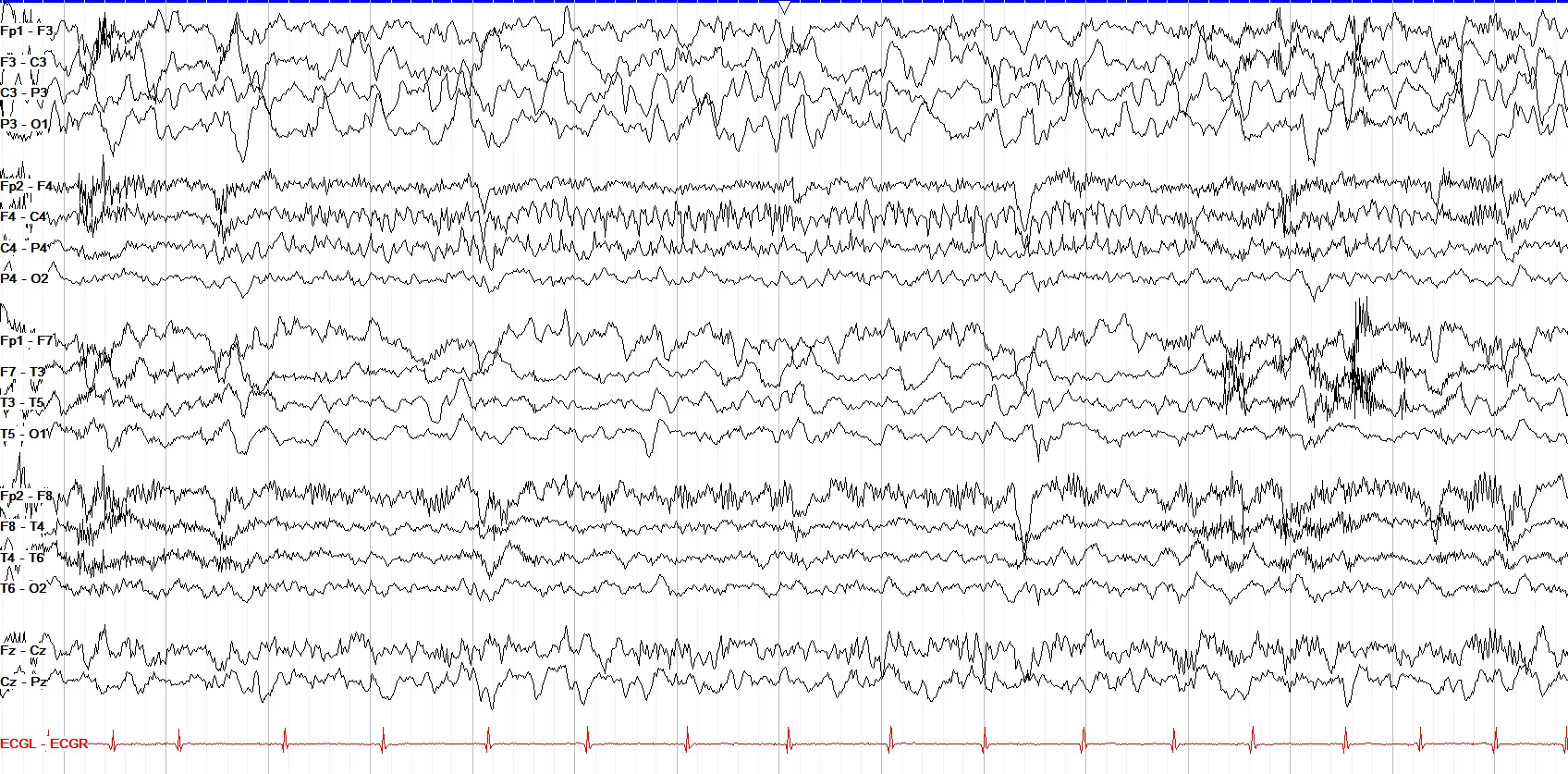
This page shows prominent high amplitude, sharply contoured delta slowing throughout the left hemisphere, more prominent in the parasagittal than temporal chain. This is consistent with an underlying focal dysfunction of that region and breach artifact. There is mu rhythm seen in the right parasagittal region, marked by arch-like alpha activity that’s often sharply contoured.
Wicket Waves
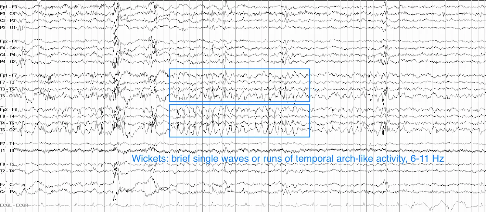
Wicket waves are so named because their arch-like shape resembles a croquet wicket (the hoop planted in the ground that a ball is hit through). Somewhat similar to mu, wickets are alpha range (again, usually 7-11 Hz) waveforms often in very brief runs; unlike mu, however, wickets are seen in the temporal regions and tend to be seen more in the drowsy state.
Don’t confuse wickets for epileptiform discharges (even when some wickets may be larger in amplitude than others) as wickets have no aftergoing slow wave, do not disturb the background, and are non-evolving even though they may come in runs.
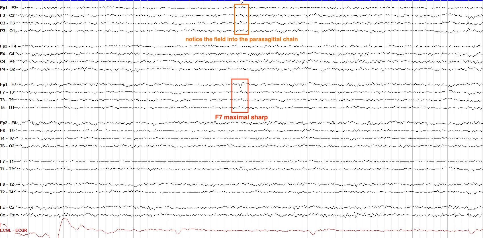
This discharge is in the left temporal region, where wickets are expected to be seen. However, this particular discharge is in isolation rather than a run, has a field into the left parasagittal chain, and rather than the classic arch-like morphology of a wicket it shows a spike and slow wave appearance that is more in line with an epileptiform discharge.
Rhythmic Mid-Temporal Theta of Drowsiness (RMTD)
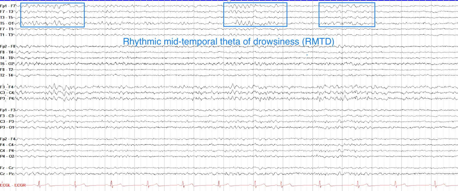
Another example of EEG having very aptly named findings, rhythmic mid-temporal theta of drowsiness (RMTD) is seen as sharply contoured, rhythmic theta activity restricted to the temporal regions, usually lasting a few seconds without evolution. RMTD is usually most prominent mid-temporally and can be bilateral or independently unilateral. The lack of evolution of RMTD (meaning its frequency and amplitude do not change over its duration) helps to differentiate it from temporal seizures or brief ictal rhythmic discharges (BIRDs).
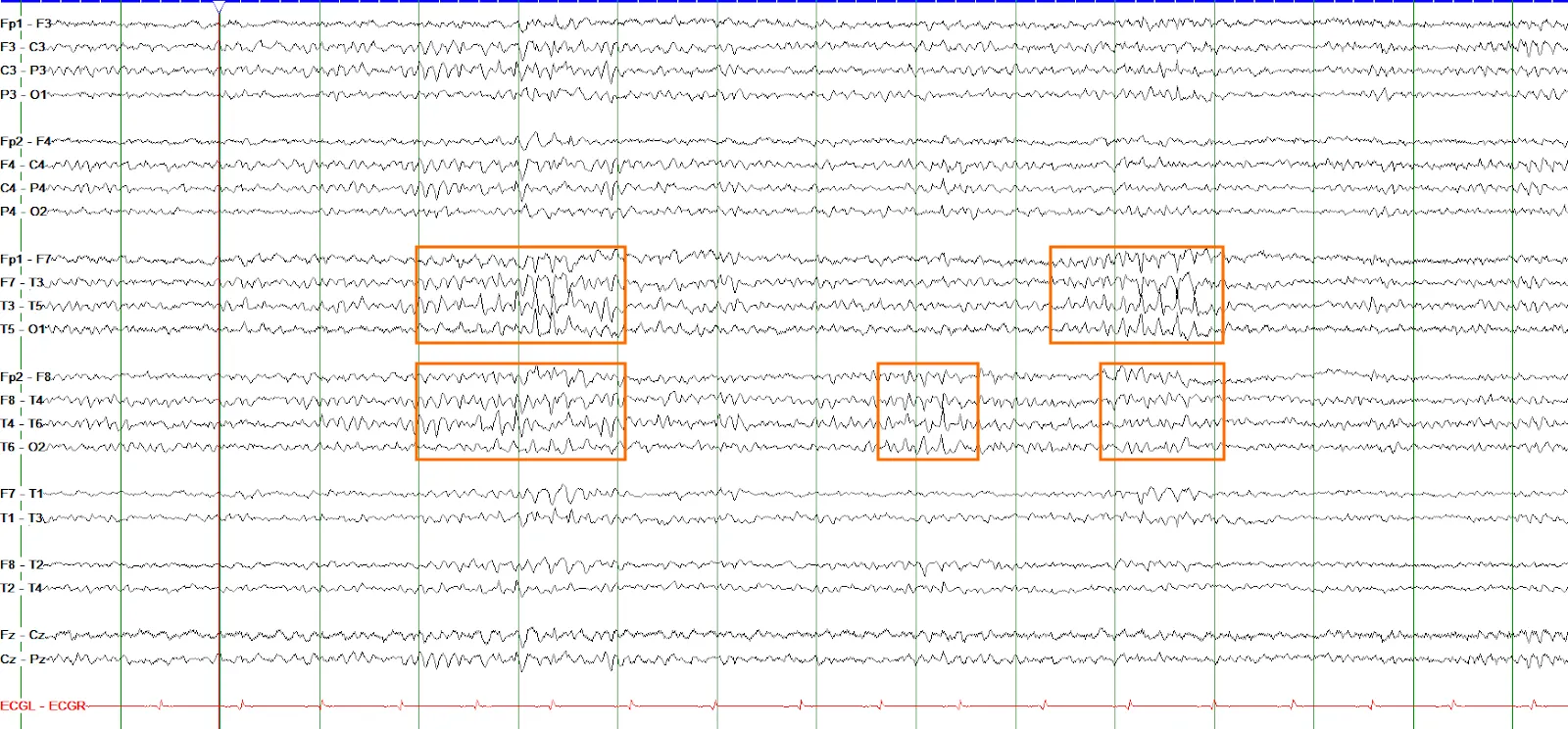
These waveforms are sharply contoured, somewhat arcuate in appearance, come in bursts of about a second, are non-evolving, and do not have an aftergoing slow wave. All of these characteristics are consistent with wicket waves. Note that these wickets are present bilaterally and unilaterally, which is expected.
Lambda Waves
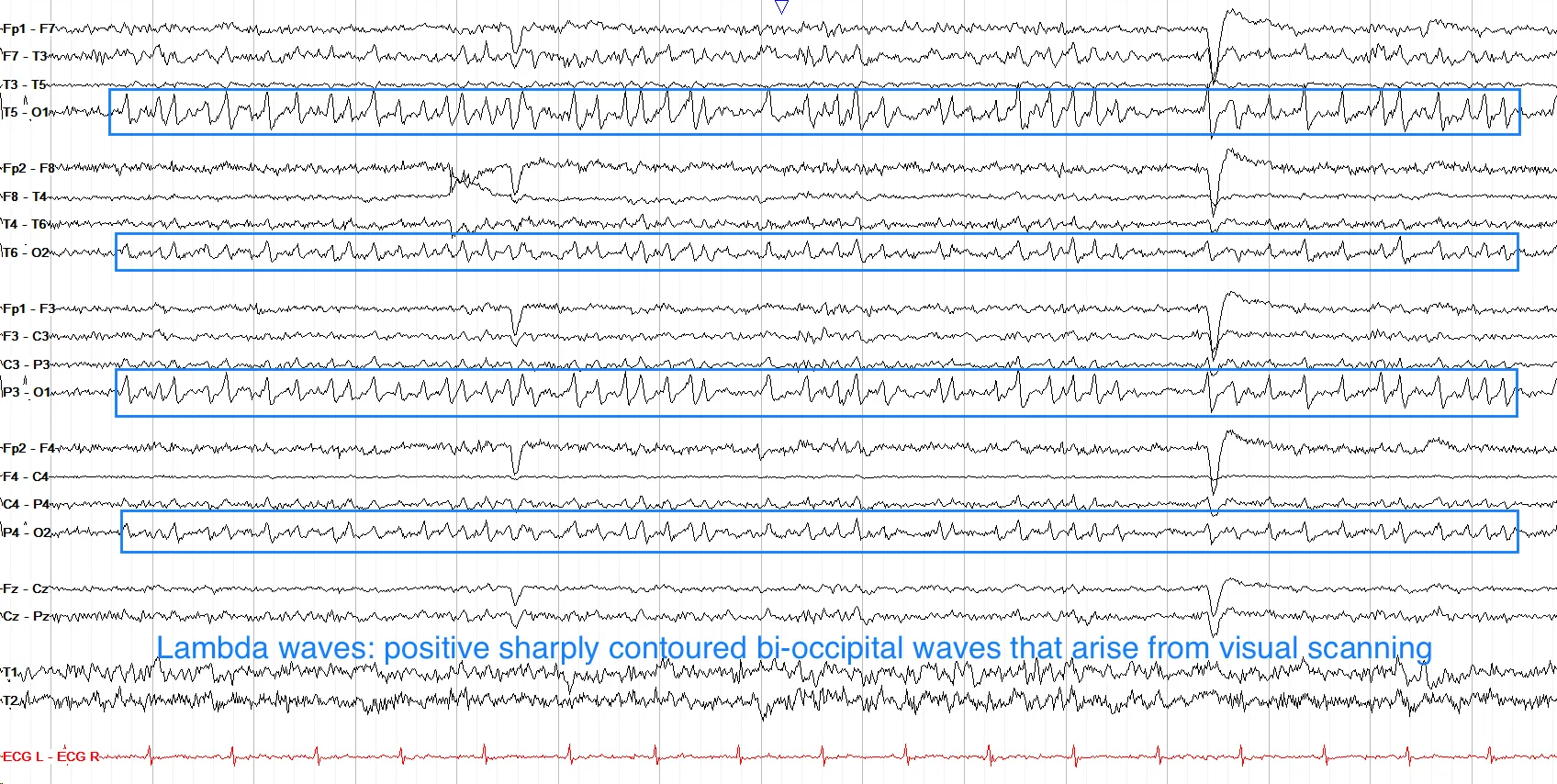
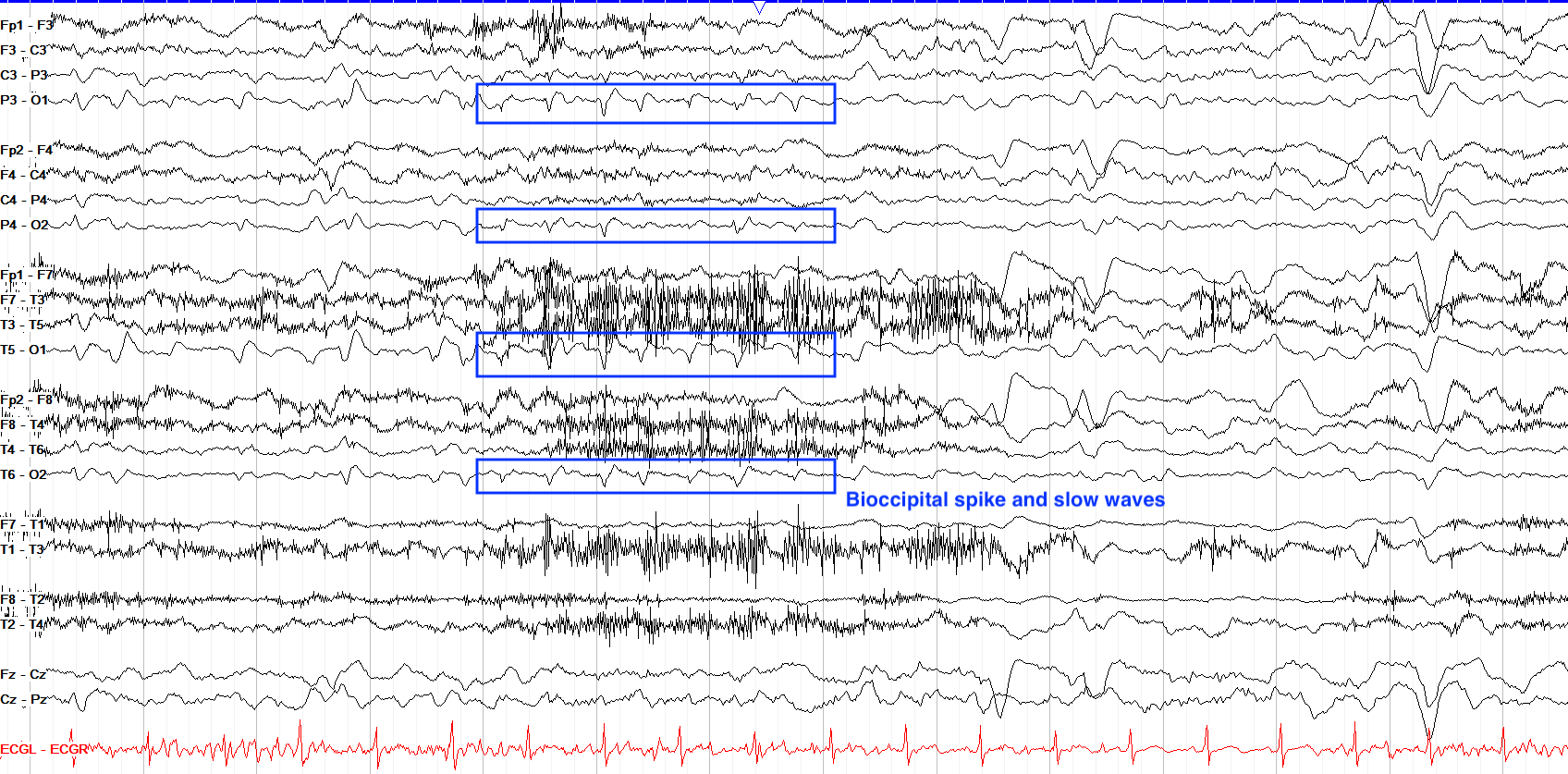
Lambda waves emerge in the awake state when one is visually scanning something, such as with reading. Lambda waves are bilateral, symmetric sharply contoured positive occipital waves with a sail-like appearance, very similar to POSTS; unlike POSTS, however, lambdas come with other evidence of wakefulness—such as eye blinks, myogenic artifact and more—rather than early sleep.
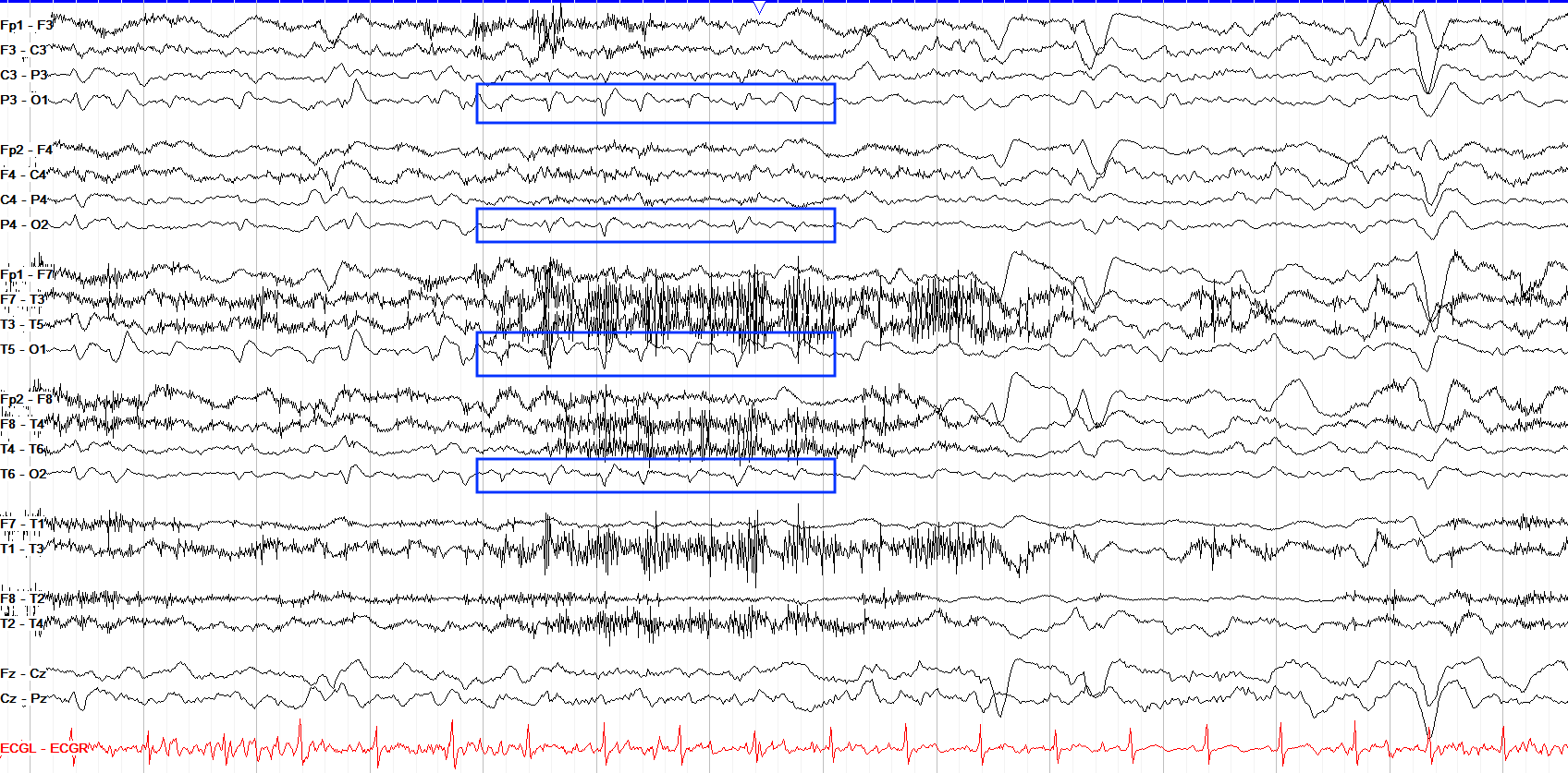
The marked waveforms, while relatively symmetric as you’d expect with lambda waves, are otherwise wholly different. First off, they have a negative polarity while lambdas are positive. Second, the marked waves have the classic epileptiform morphology of a spike and slow wave, although some show this more clearly than others. Don’t let them fool you just because there are a series of them; epileptiform discharges can come in nonevolving runs as you see here.
Benign Epileptiform Transients of Sleep (BETS)
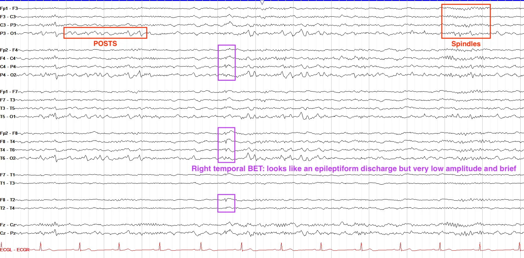
Benign epileptiform transients of sleep (BETS) are also called small sharp spikes (SSS). While BETS have a slightly oxymoronic name, they are in fact a normal, benign finding that you can see in the drowsy and asleep states. In morphology they are very similar to epileptiform spikes, including having a field, but by definition BETS are low amplitude (technically less than 50µV) and short duration, and they characteristically lack an aftergoing slow wave—a key feature that helps distinguish them from true epileptiform discharges. They can be seen most often in the temporal chains.
Differentiating BETS from real epileptiform discharges can be tricky; a good rule of thumb is that if the discharge is very low amplitude and seen only once or a few times, and only during sleep, it’s probably a BET, though you should make note of it and keep a look out for anything more suspicious.
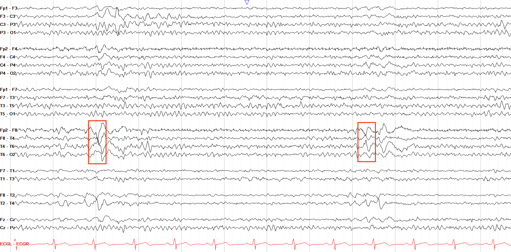
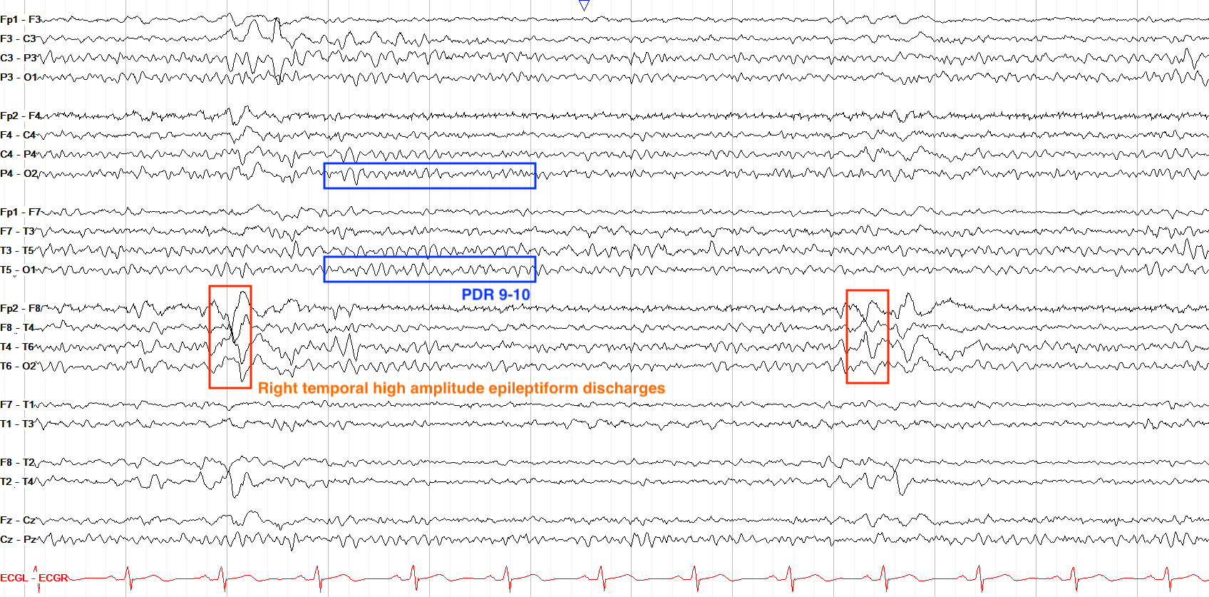
This is actually an easy question, as you can see a clear PDR of 9-10 throughout this page. Therefore, the marked discharges cannot be BETS, as BETS are only present in sleep by definition. In looking at the marked waveforms, however, they are also too high amplitude and last a little too long to be BETS, which should be very small (<50µV) and short.
14 and 6 Positive Spikes
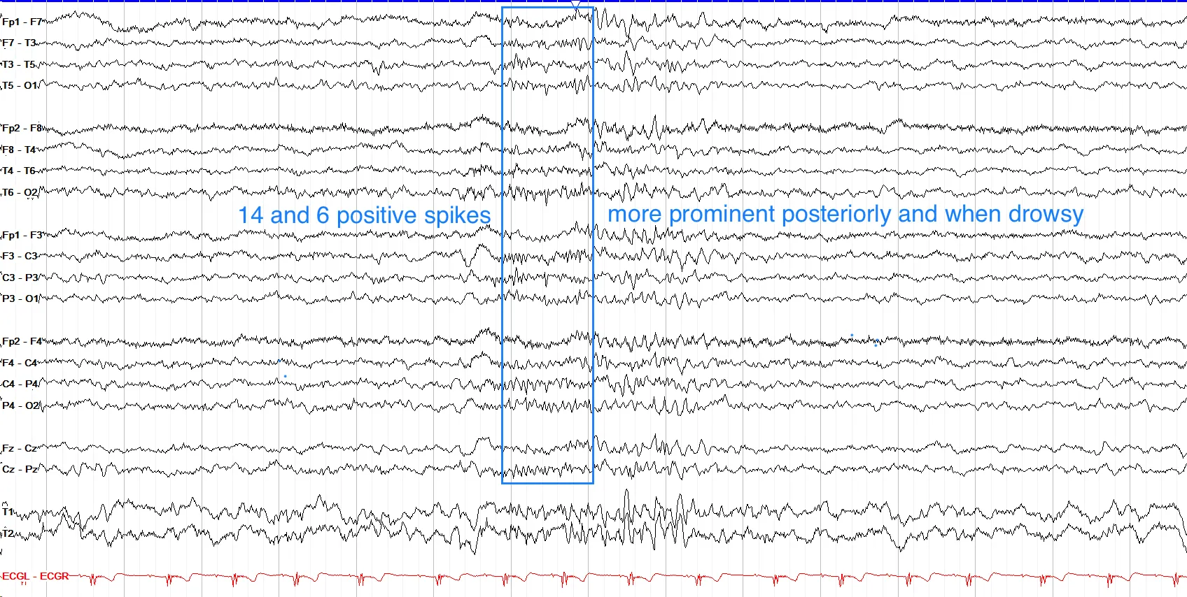
Taking the crown for perhaps the most uninspired name of EEG findings, 14 and 6 positive spikes describe 1-2 second bursts of sharply contoured positive waveforms that come in frequencies of either 14 Hz (13-17 Hz) or 6 Hz (5-7 Hz), or an admixture of both. They should be bilateral and synchronous, and are more common in the posterior quadrants when drowsy. They are most often seen in young adults and adolescents, and are not associated with increased epilepsy risk. In this first example below, you can see a one second run of diffuse ~14 Hz activity amongst a background suggestive of drowsiness, consistent with 14 Hz positive spikes.
In this second example, you can see a one second run of diffuse ~5-6 Hz activity, consistent with 6 Hz positive spikes.
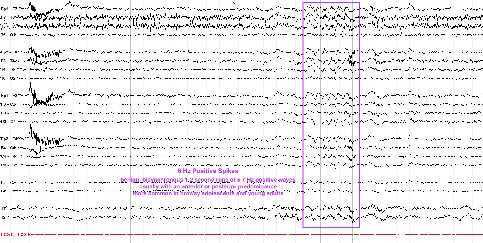
There is a similar normal variant that shouldn’t be confused with 14 and 6 positive spikes, termed 6 Hz phantom spike and waves. While 14 and 6 positive spikes are comb-like without any slow waves, 6 Hz phantom spike and waves are comprised of 5-7 Hz brief runs of very low amplitude spikes followed by an equally tiny slow wave. These are often generalized but have two subtypes: WHAM (waking, high amplitude, anterior, male) or FOLD (female, occipital, low amplitude, drowsy). These subtypes describe themselves, really, and the important thing to remember is that despite their having “miniature” epileptiform morphology these discharges are not indicative of seizure risk, though there is some data that WHAM may be associated with risk for epilepsy while FOLD are not.
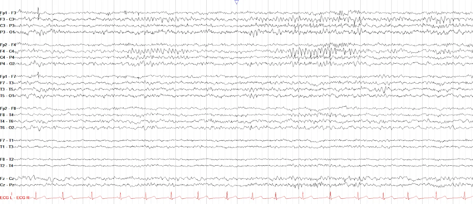
This page has intermittent periods of bilateral sharply contoured, arch-like alpha activity over the parasagittal regions without associated disturbance of the background. This is consistent with mu rhythm, the idling activity of the sensorimotor cortex that goes away when you think about movement or actually move an extremity.
- Mu is the idling activity of the sensorimotor cortex, seen as arch-like alpha over the centroparietal regions; it is suppressed by thinking about or initiating movement.
- Wicket waves are arch-like, alpha range waveforms seen in the temporal regions, most often when drowsy; they lack an aftergoing slow wave, do not disturb the background, and are non-evolving.
- Rhythmic mid-temporal theta of drowsiness (RMTD) is sharply contoured rhythmic theta in the mid-temporal regions lasting a few seconds without evolution.
- Lambda waves are bilateral, symmetric, positive occipital waves with a sail-like appearance caused by visual scanning when awake (they look similar to stage I sleep POSTS).
- Benign epileptiform transients of sleep (BETS), or small sharp spikes (SSS), are low amplitude very brief transients with no aftergoing slow wave seen usually temporally in the drowsy and asleep states.
- 14 and 6 positive spikes are 1-2 second bursts of sharply contoured and posterior predominant bilateral positive waveforms at 14 Hz or 6 Hz, usually seen when drowsy.