Pediatric EEG Evolves into the Adult
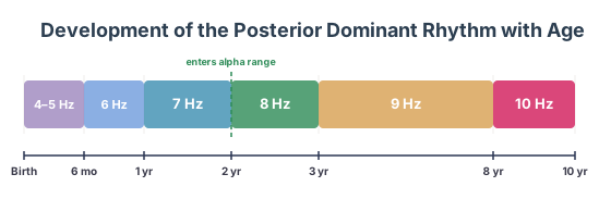
After the first month of life, a baby graduates from being a neonate to an infant, and with that moves from the neonatal montage to the full standard 10-20 montage. Just as with neonates, though, infants and children have an evolving EEG that requires a solid understanding of the normal timeline for proper interpretation. A key part of the pediatric EEG is its evolving PDR, discussed in detail below, but summarized as follows:
Infancy
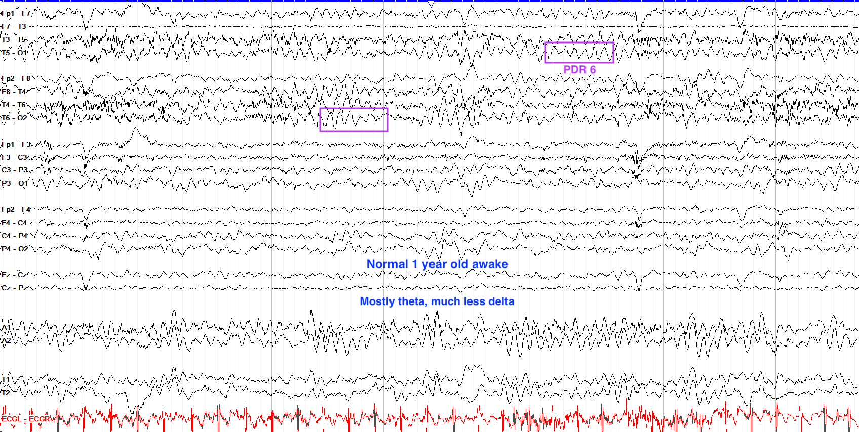
The first year of life on EEG is characterized by a preponderance of slow, high amplitude delta activity. The recording should be continuous and symmetric, but not particularly reactive to eye opening until 2-4 months. By 6 months a PDR of 4-5 Hz starts to emerge, and though delta still predominates the background that gradually transitions to theta predominance during wakefulness by 12 months. In drowsiness, though, the background often remains slower and higher amplitude, with a lot of 1-2 Hz activity up to 200uV, particularly early in that first year of life. By 1 year of age, the expected PDR is 6 Hz.
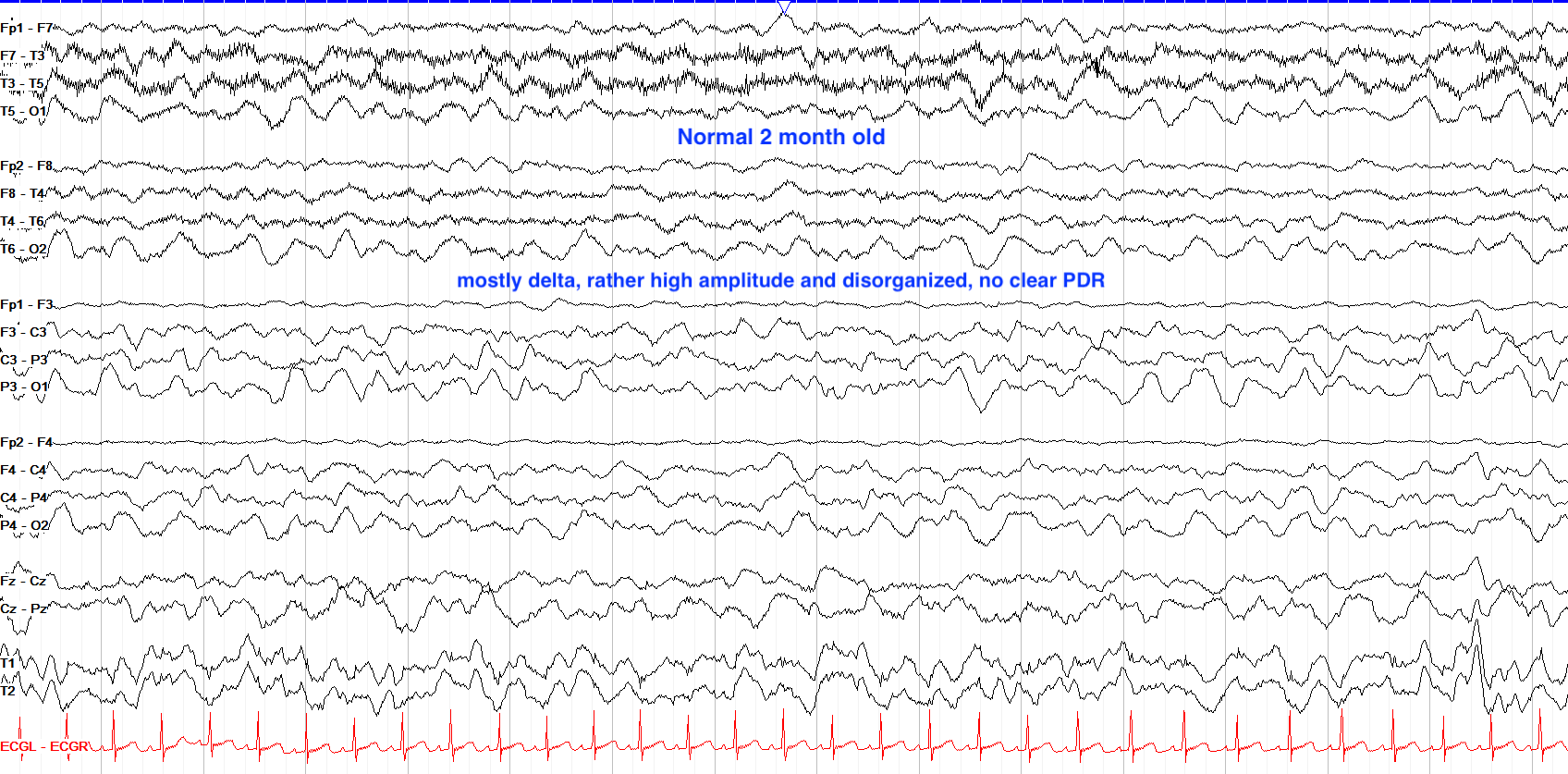
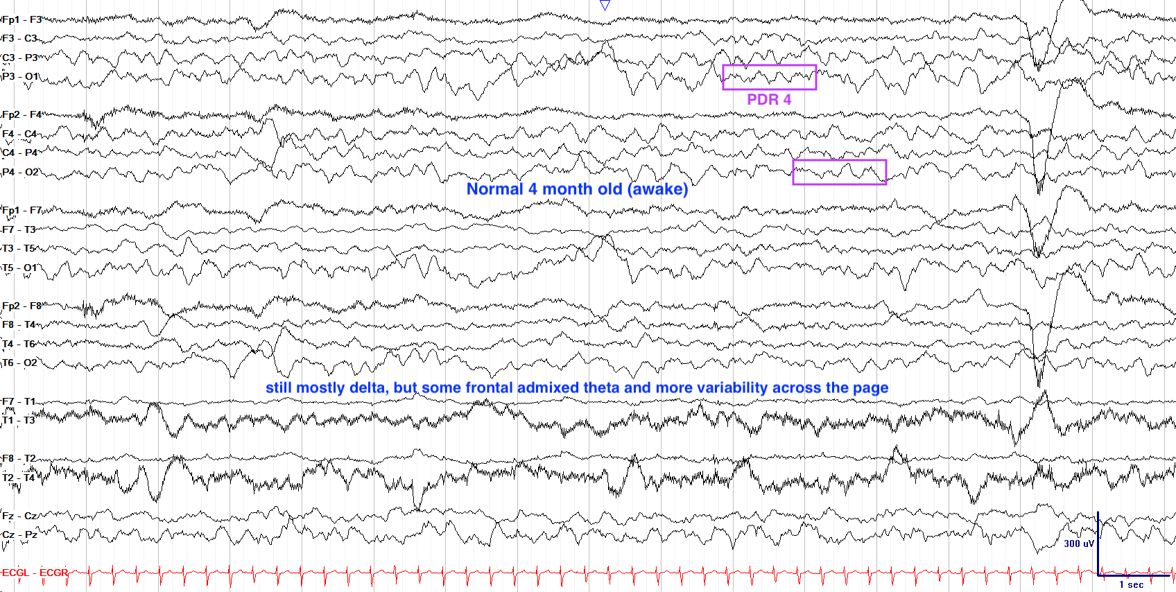
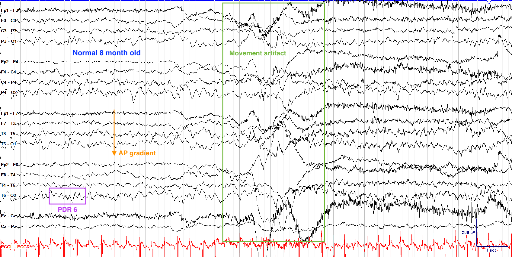
Below are a few examples of normal waking tracings at various time points for infants; note the general trend, over time, of less delta and more theta, and a better formed anterior-posterior gradient. These tracings have their sensitivities lowered from the standing 7uV/mm to between 10-20uV/mm for clarity as, generally, pediatric tracings are higher amplitude than adults.
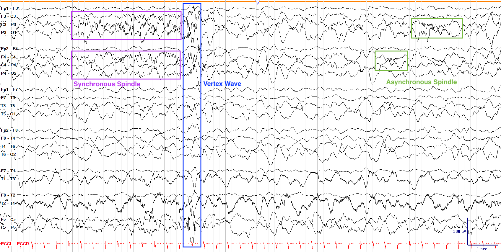
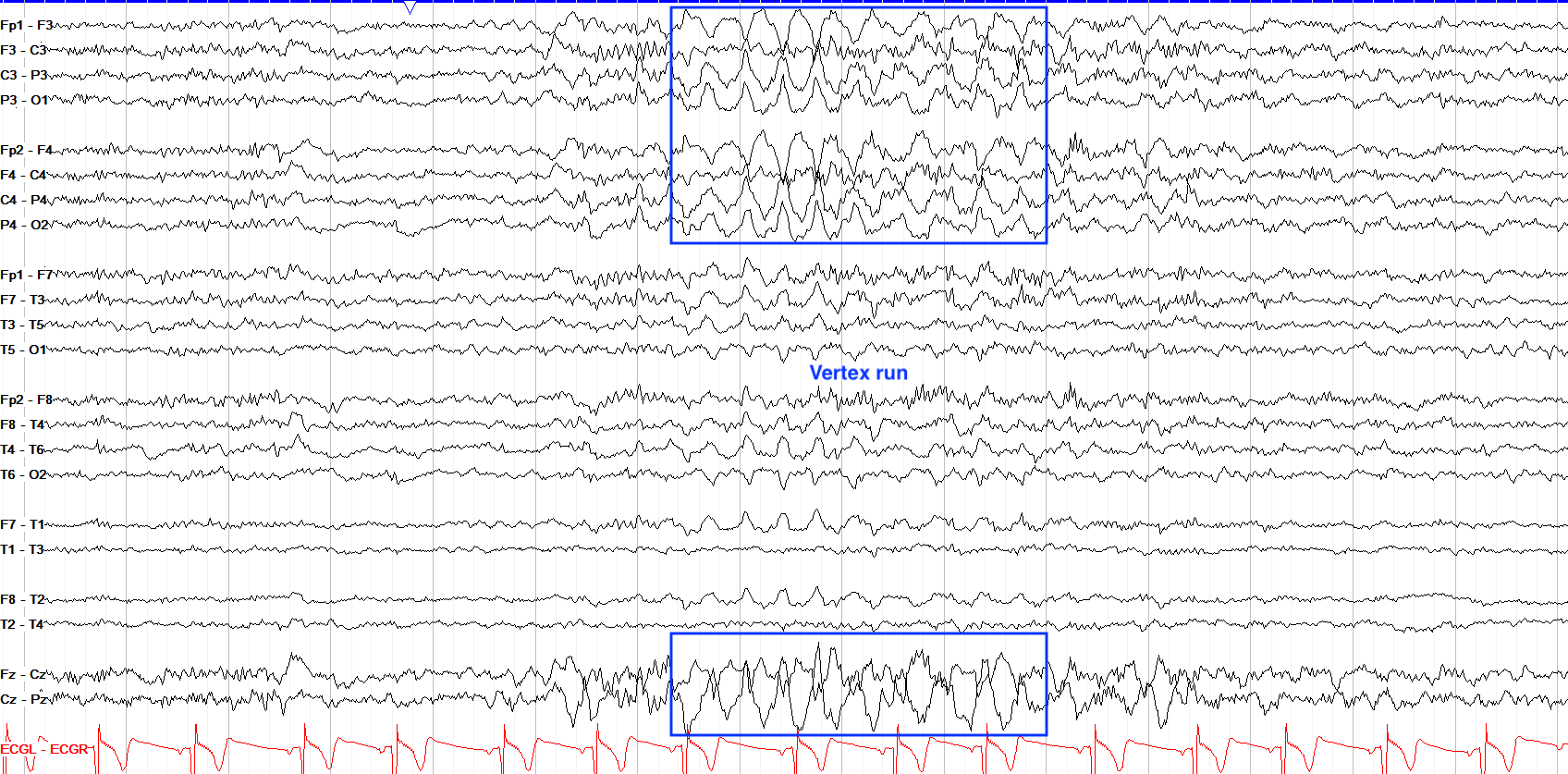
Regarding sleep, by 2 months of age stage II sleep spindles develop; initially they can be very prolonged (up to 15 seconds at a time), and can remain asynchronous till up to 2 years of age. Along with these spindles other sleep architecture arises including vertex waves in stage I and K complexes in stage II; these formations can be extremely prominent in children, with vertex waves, in particular, often looking very sharp and coming in long runs, so don't confuse them for B(I)RDs or seizures.
In the first months of life, up to half a baby's sleep time can be REM sleep, but this proportion decreases to about a third by 1-2 years of age. The example below is from a 4 month old healthy baby; note the high amplitude vertex wave, rather prolonged spindle in the first half of the page, and the asynchronous and poorly formed spindle near the end of the page.
You won't often try activation techniques in babies, but prolonged crying can cause a hyperventilation response, and if you do attempt photic stimulation, a slow driving response of 1-3 Hz is seen starting around 6 months of age.
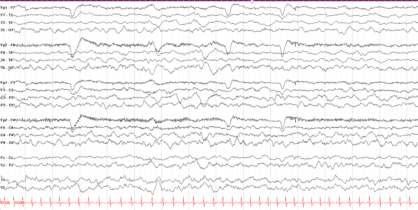
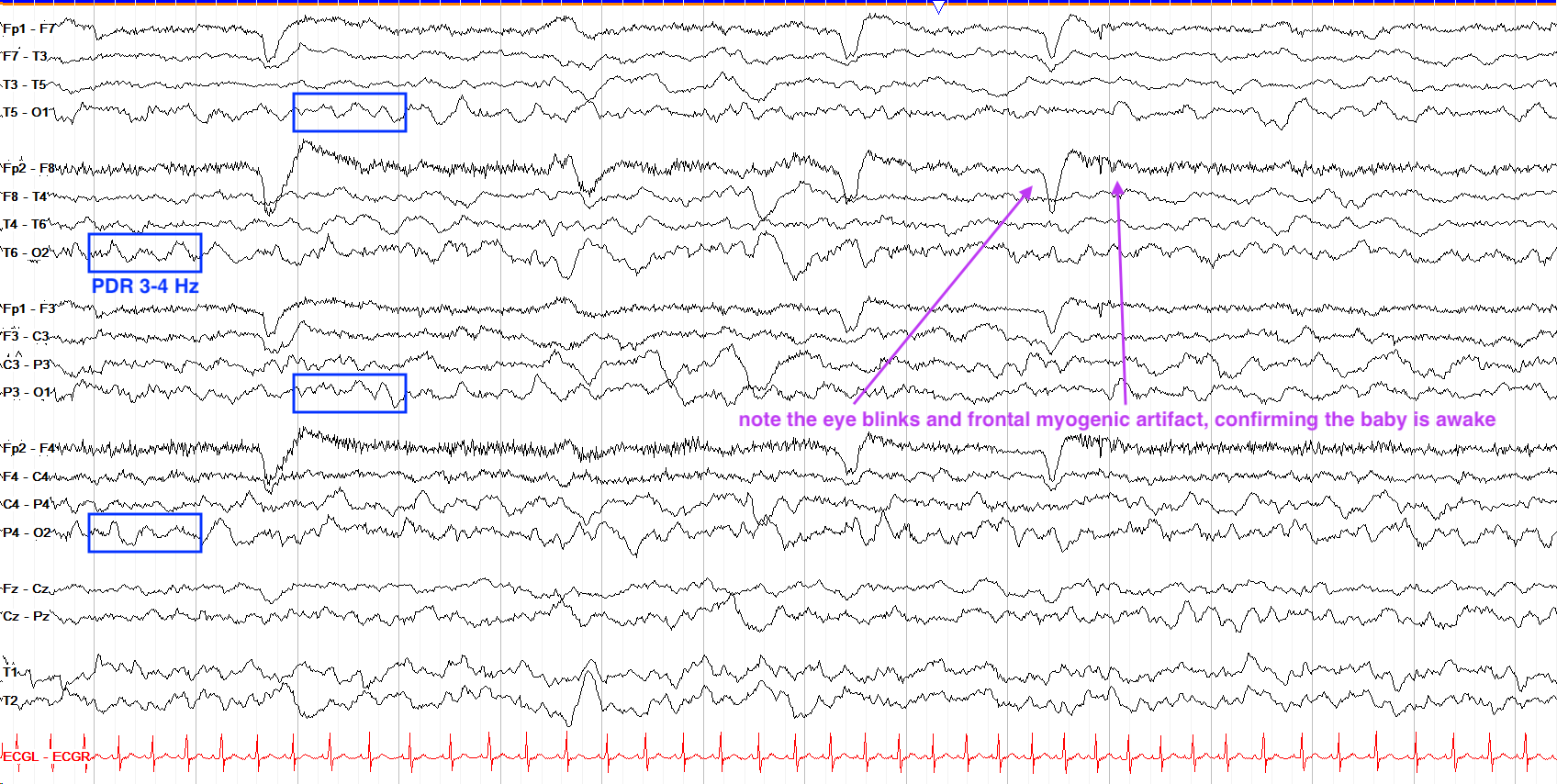
First of all note that this tracing is in the awake state, given multiple eye blinks across the page. This tracing shows a PDR of 3-4 Hz, which would be normal for a 2 or 3 month old awake baby, and maybe even the lower limit of normal for a 6 month old (their normal is 4-5 Hz), but by 1 year of age you should be seeing an awake PDR of at least 6 Hz.
Early Years
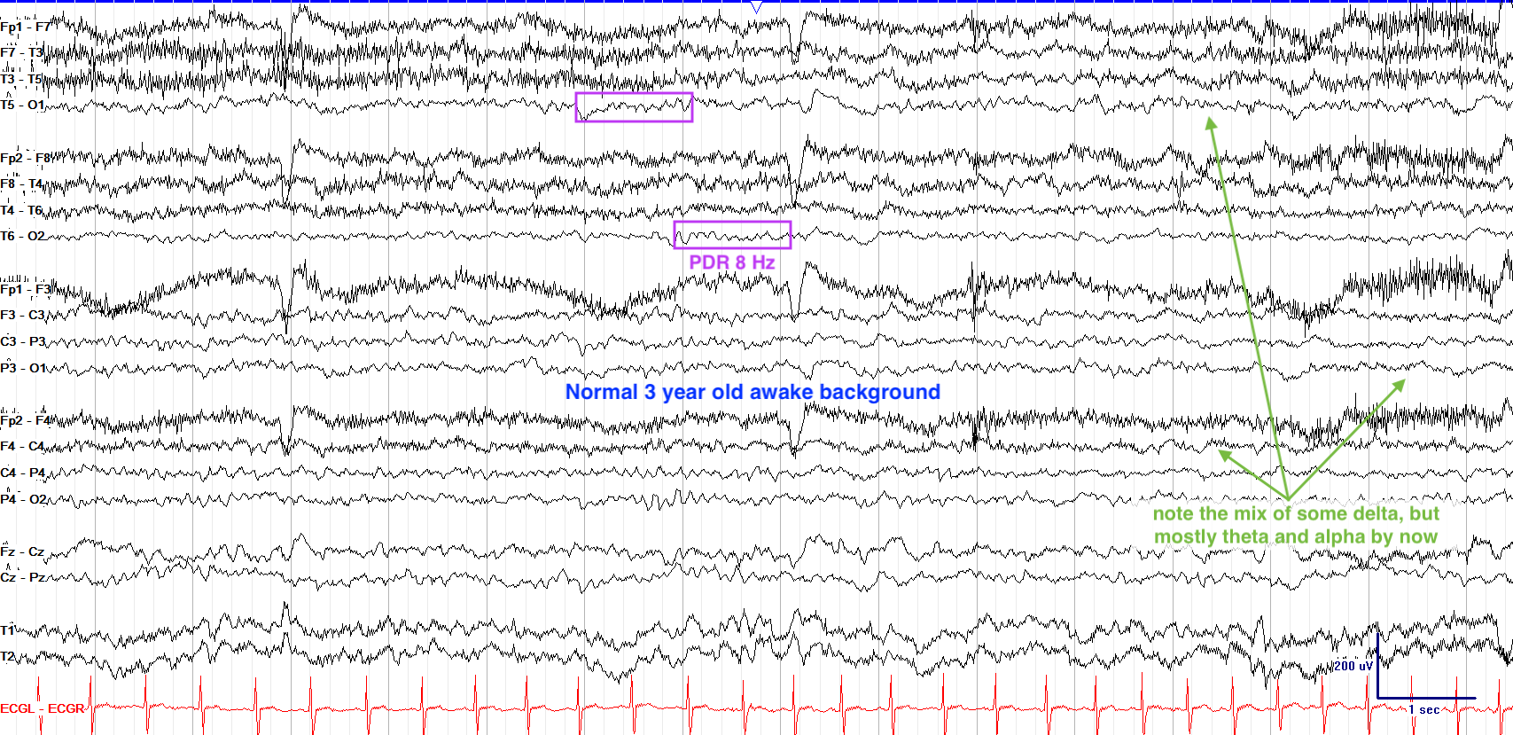
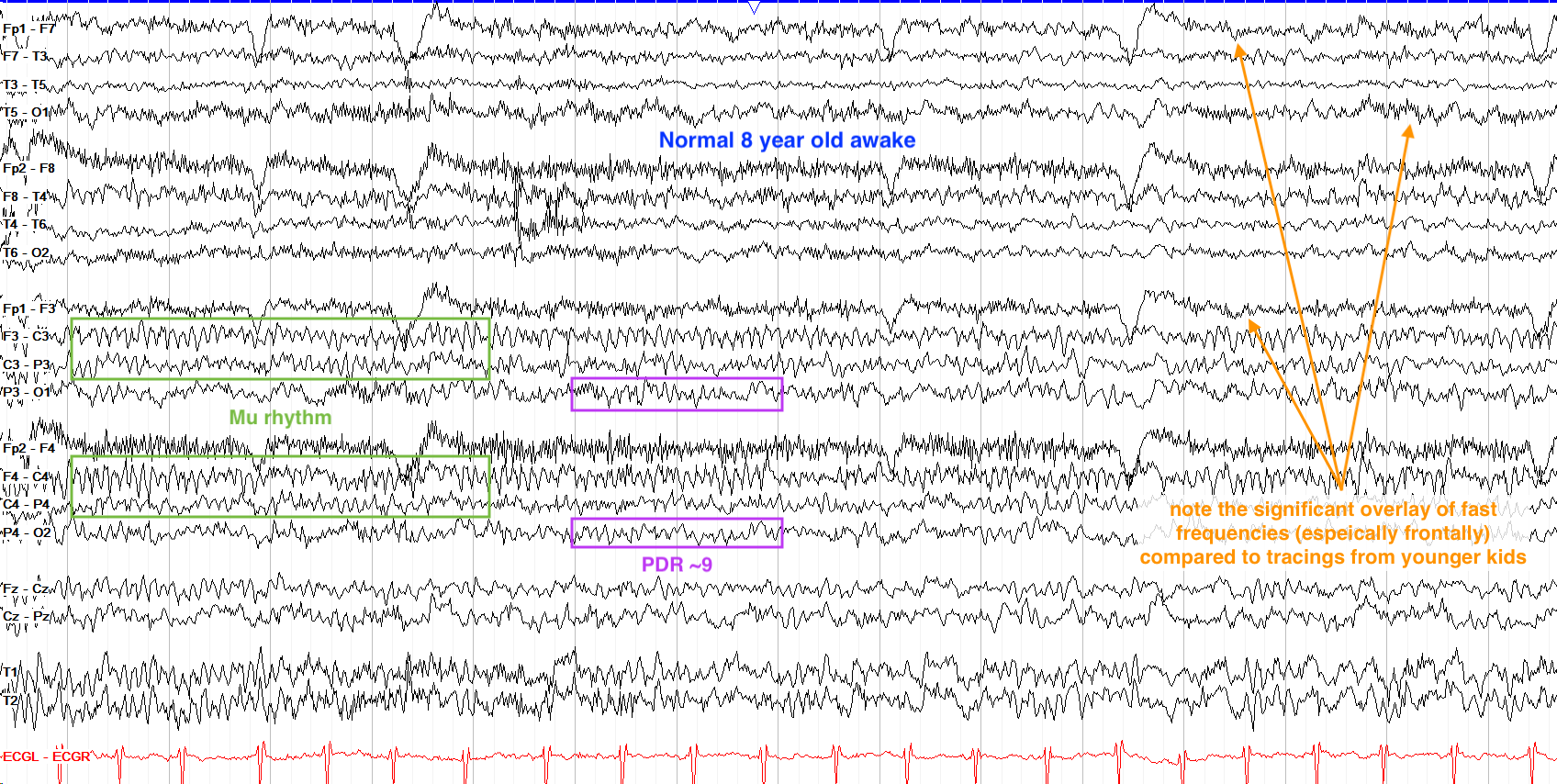
After the first year of life, most of the delta activity seen in infants' waking background shifts to theta and further on to alpha by now. The PDR continues to evolve, reaching 8 Hz by 3 years of age, and remains higher voltage than you'll see in adults. You'll also start to see some of the same benign variants and rhythms from adult tracings, including lambda waves with visual scanning and the mu rhythm as the idling activity of the sensorimotor cortex. Sleep architecture remains very high amplitude, especially slow wave sleep.
In the set of examples below, focus on the overall progression of the background, as it moves from a mixture of theta and alpha at 3 years to much more alpha and beta (similar to adult tracings) by 8 years of age. Again, note that these tracings have a lowered sensitivity (10uV/mm) to allow for better clarity.
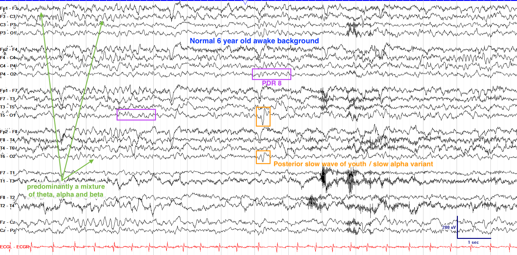
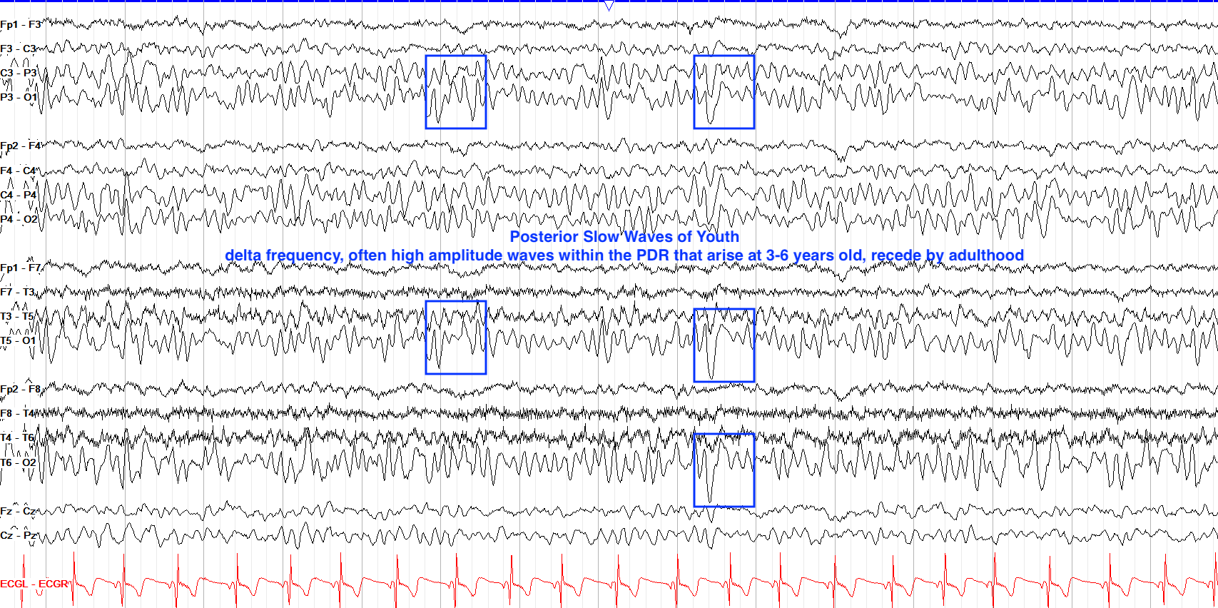
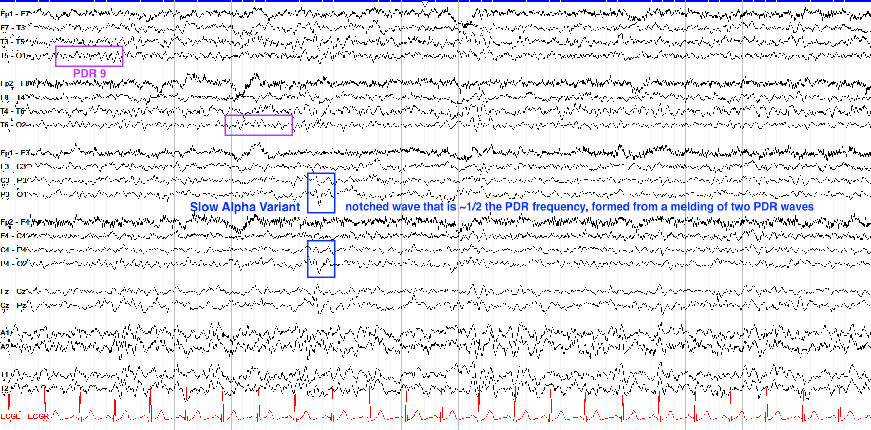
From 3-6 years of age, a new waveform also arises, posterior slow waves of youth. These are rather high amplitude, spike-like waveforms that exist within the PDR and similarly attenuate with eye opening; they are often but not always bilateral, and can predominate on one side or the other. Similarly, there can emerge slow alpha variants, which are essentially two waves from the PDR combining into one, which is thus about half the frequency of the usual PDR. Below are examples from a 5 year old patient, with a PDR of 9 Hz (normal for age). Note how the posterior slow wave of youth has a delta frequency but is surrounded by the PDR without really interrupting the PDR's pattern or background otherwise, and how the slow alpha variant has a notched appearance that should not be mistaken for an epileptiform discharge.
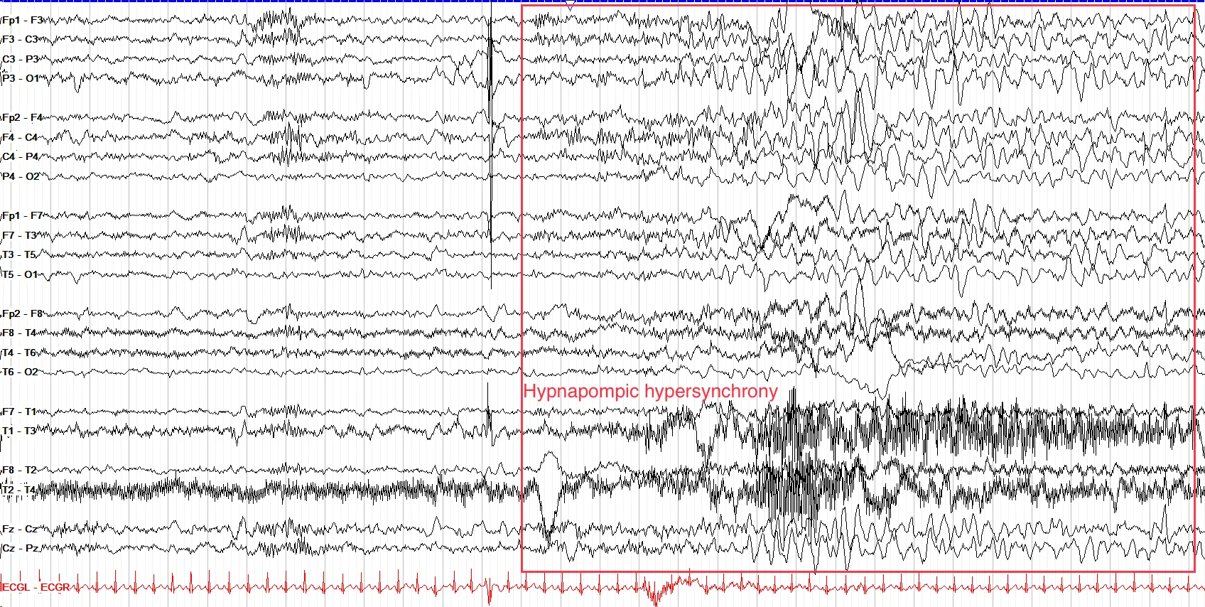
Note that drowsiness in children may not be accompanied by the classic slow, roving eye movements seen in adults. Throughout this time period, sleep architecture should be synchronous and essentially in line with what you see in adults, although generally more prominent and high amplitude, with vertex waves often coming in runs. You can also see hypnagogic or hypnopompic hypersynchrony, in which high amplitude, synchronized slow waves arise in the transitions between waking and sleeping states.
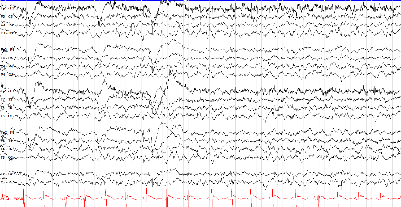
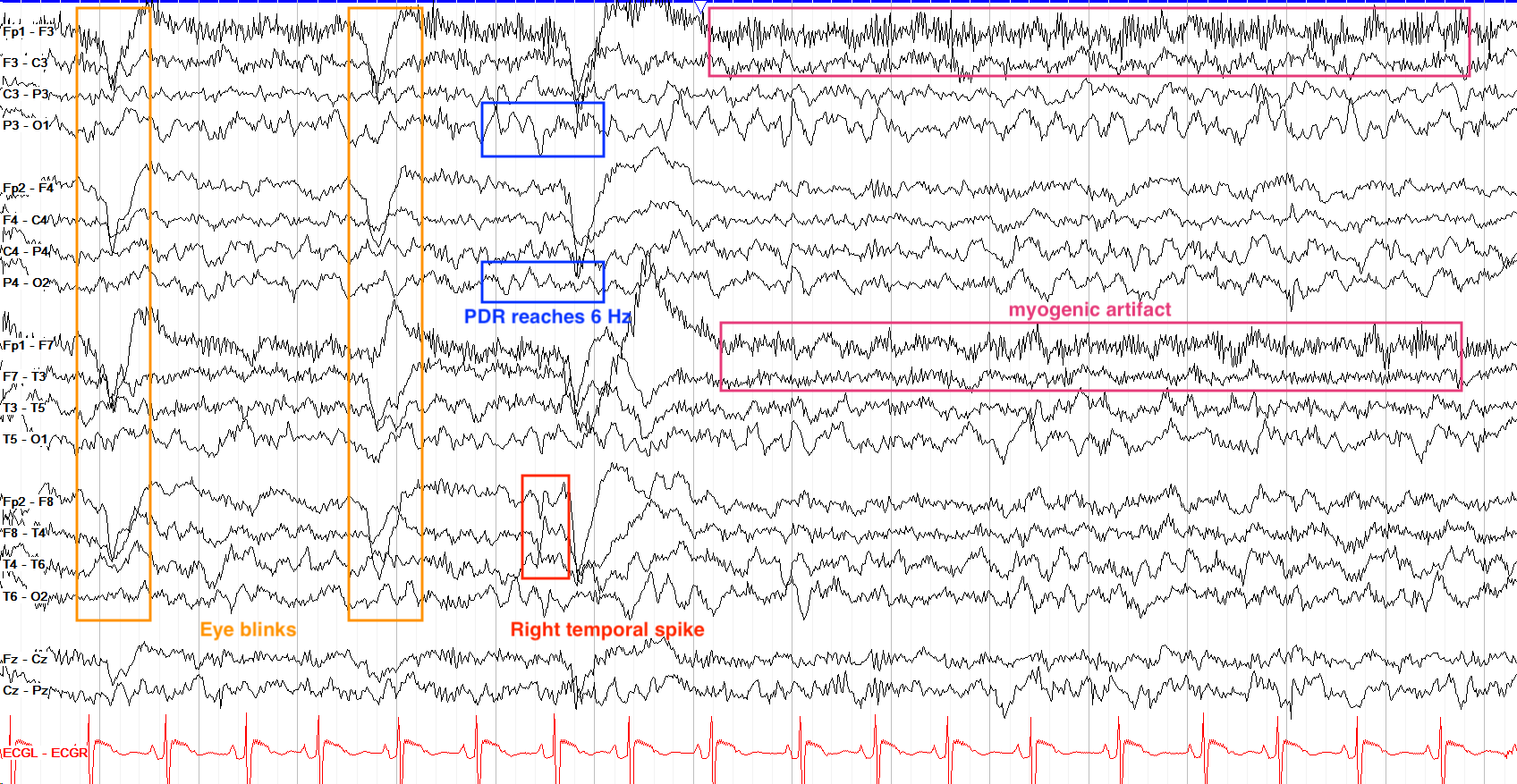
First of all, we know this patient is awake because we see eye blinks and a lot of frontal myogenic artifact. Second of all, this tracing has a lot of delta and theta activity, which might be normal for a very drowsy 7 year old, but in an awake patient of this age we expect to see much more alpha activity with a PDR of at least 8-9 Hz. Here, though, the PDR doesn't get past 6 Hz, and that plus the excess delta and theta activity merits a call of mild generalized slowing. There is also a right temporal spike, although this patient has multifocal spikes seen elsewhere in the tracing too.
Adolescence to Adulthood
By the time kids reach adolescence, their EEG looks quite similar to adults, with a PDR that usually approaches 10 Hz and with a dominant mixture of alpha and beta throughout. Unlike adults, adolescents can still have some theta activity in their waking background, but this should fade away into the teen years.
In adolescence, the prominent hypnagogic and hypnopompic hypersynchrony of the early years recedes, and as teens grow into adults they lose posterior slow waves of youth. Sleep architecture remains quite prominent in teenagers and young adults, with vertexes continuing to often come in runs even into some patient's thirties; the example below is from a 15 year old patient.
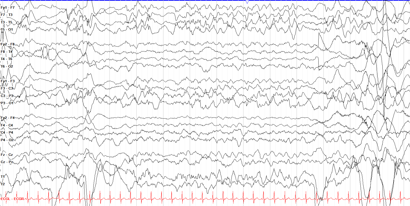
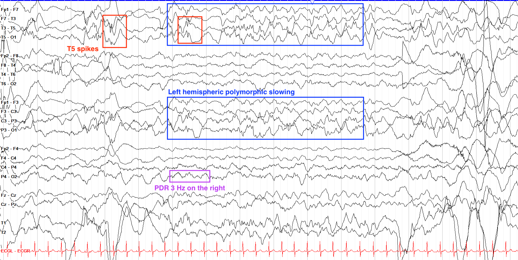
Recall that by 6 months of age a baby should have a PDR of 4-5 Hz. Here, we see on the right side a PDR of about 3 Hz, which is age appropriate. However, on the left side there is broad hemispheric polymorphic delta slowing, suggestive of an underlying structural abnormality. On top of that, we see a few left temporal spikes, phase reversing at T5 suggesting focal cortical hyperexcitability from that area. The high amplitude, messy activity toward the end of the page is mostly movement artifact.
Pediatric Syndromic EEG Patterns
From infancy into adulthood, there are a number of classic EEG patterns and syndromes that you should be familiar with, because each has different treatment requirements and prognoses. Here is an overview on the more common ones with their particular EEG signatures.
Ohtahara Syndrome (EIDEE)
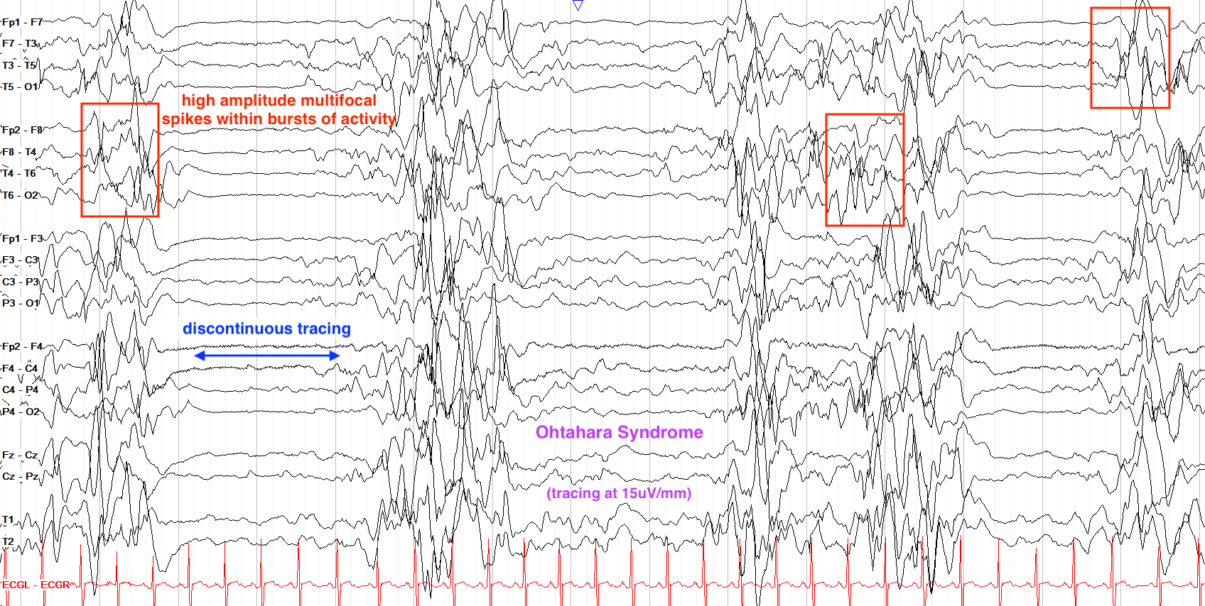
Ohtahara syndrome, now encompassed within early-infantile developmental and epileptic encephalopathy syndrome (EIDEE), is a devastating diagnosis of early infancy. On EEG, it's characterized by a burst suppression pattern with high amplitude, multifocal spikes embedded within the bursts of activity. Multiple seizure types, most often tonic seizures or spasms, are seen along with significant intellectual disability and developmental delay. EIDEE often leads to early death, but if not can progress into IESS or Lennox Gastaut Syndrome.
Hypsarrhythmia & Infantile Spasms
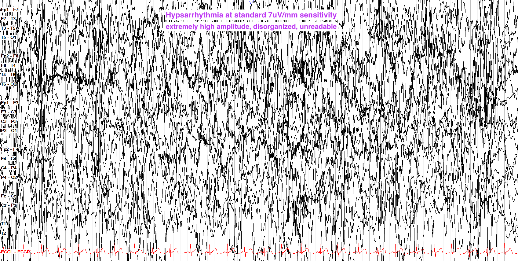
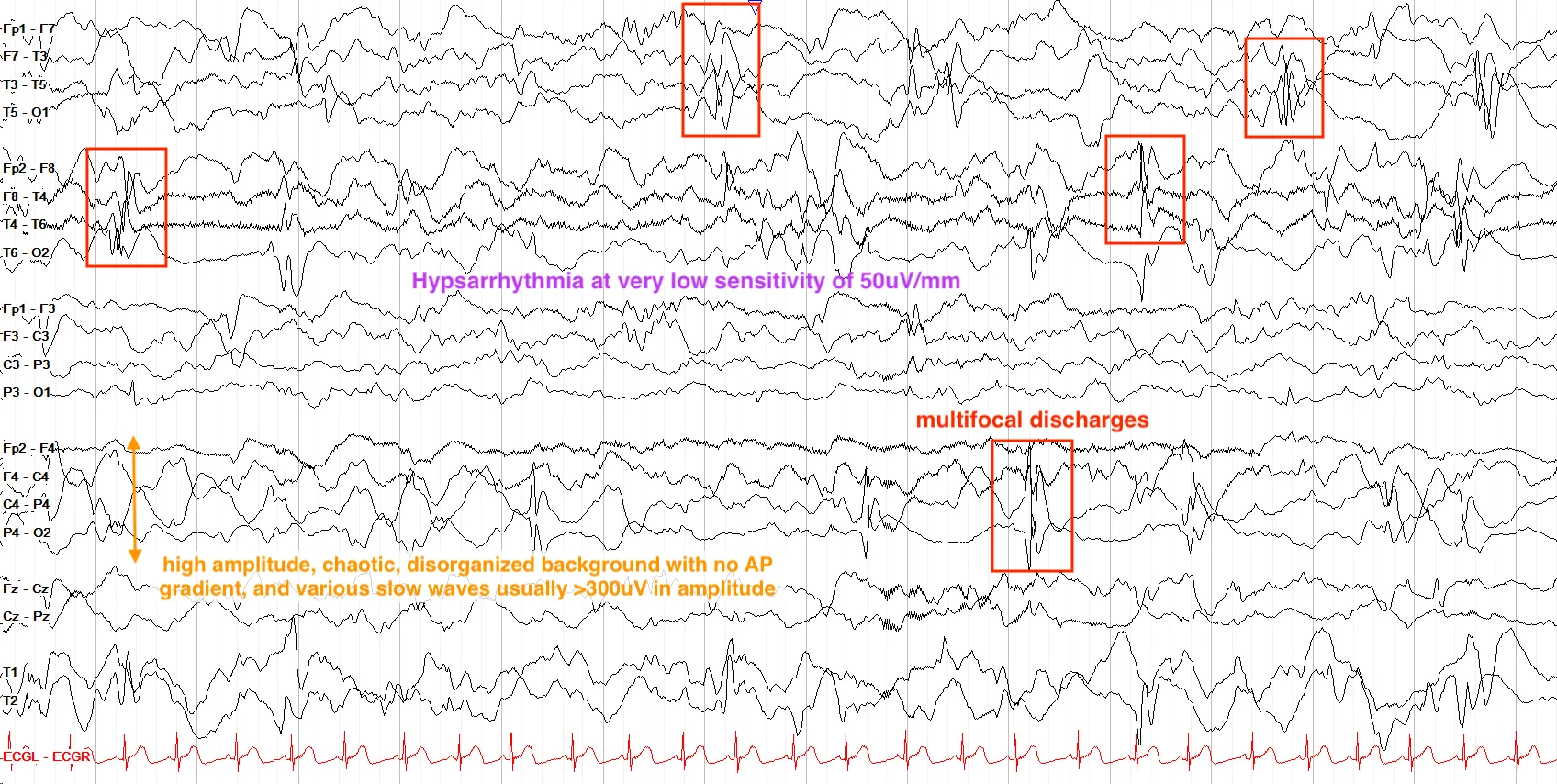
Hypsarrhythmia describes a characteristic EEG background that is extremely high amplitude, chaotic and disorganized, slow, and with multifocal epileptiform discharges throughout. As you'd expect, it's a highly concerning pattern seen most often in infants with infantile spasms, or infantile epileptic spasms syndrome (IESS, previously termed West Syndrome). This syndrome describes a triad of clinical findings including hypsarrhythmia on EEG, epileptic spasms, and developmental delay/regression. Urgent treatment is critical to prevent long term developmental sequelae and intractable seizures; if not controlled, infantile spasms can evolve into Lennox-Gastaut Syndrome.
Lennox Gastaut Syndrome (LGS)
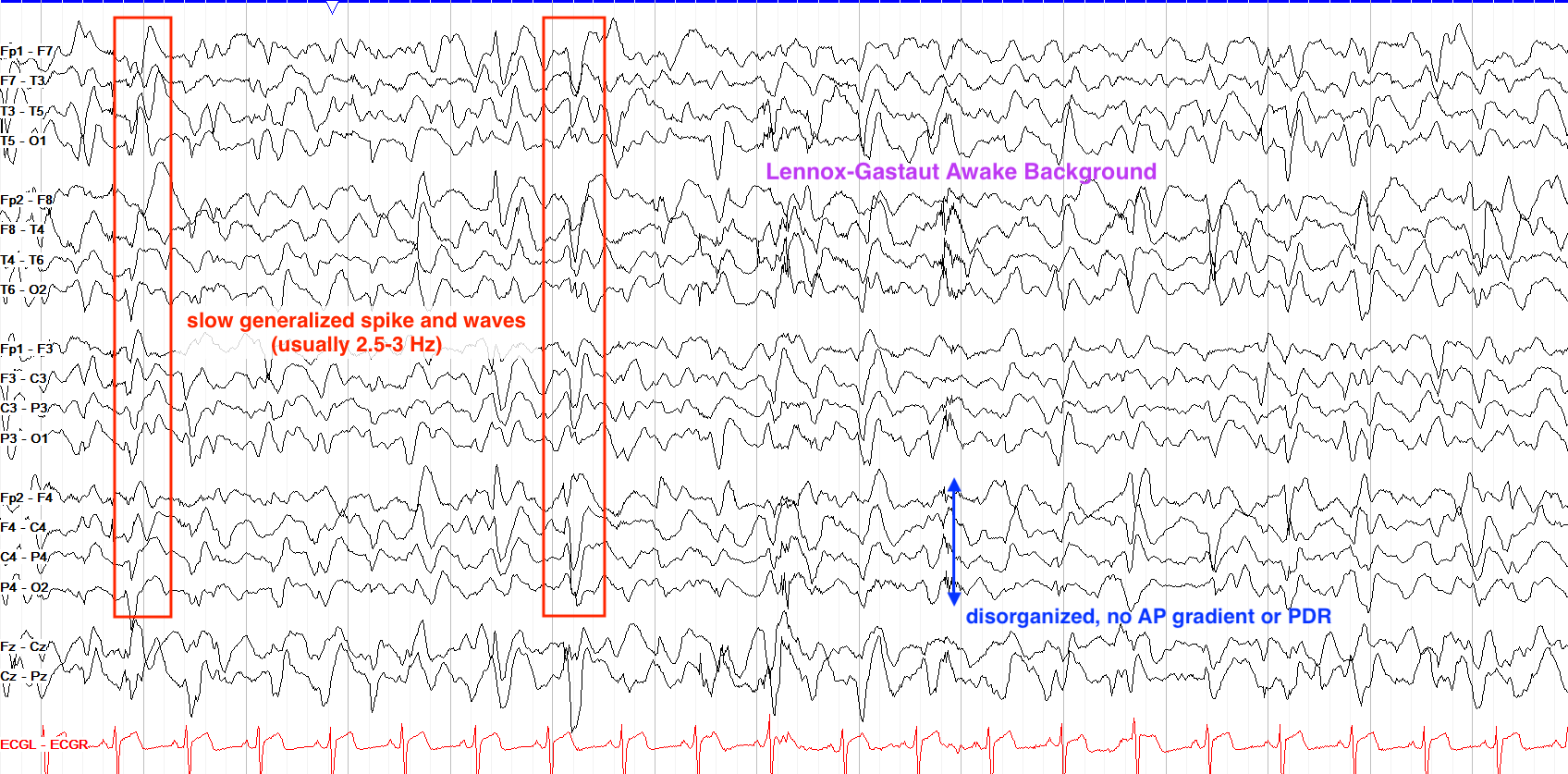
Lennox Gastaut Syndrome (LGS) can evolve from EIDEE or IESS, or arise on its own. Similar to the other two, it is a severe form of epileptic encephalopathy with multiple seizure types and intellectual impairment. The classic LGS tracing is marked by diffuse, prominent slowing and slow (typically 1.5 to 2.5 Hz) generalized spike wave discharges.
Self-limited Epilepsy with Centrotemporal Spikes (SeLECTS)
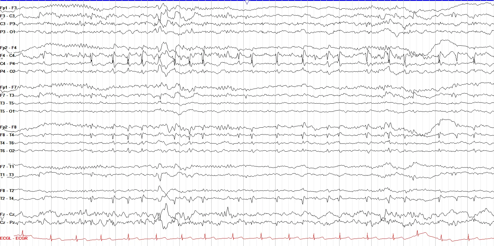
Self-limited Epilepsy with Centrotemporal Spikes (SeLECTS; previously termed benign rolandic epilepsy with centrotemporal spikes, or BECTS) is a common, descriptive and often self-limiting disorder, marked electrographically by the aforementioned centrotemporal spikes, which can be unilateral or bilateral. This condition usually emerges from 3 years of age or older, and remits by adolescence. Seizures in SeLECTS classically are focal nocturnal, and consist of unilateral facial spasms that can progress to involve the ipsilateral arm and leg.
Absence Epilepsy
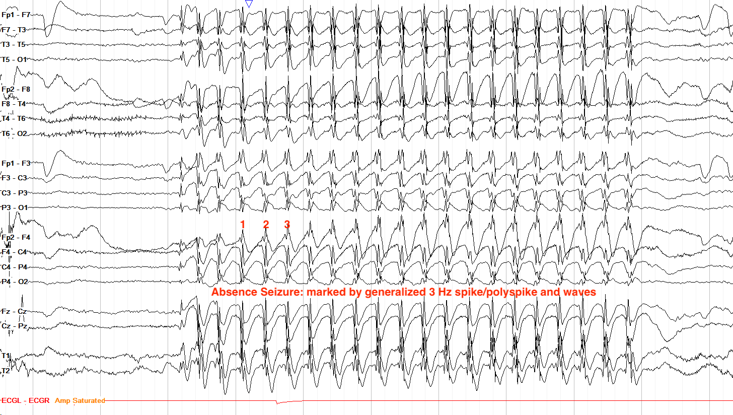
While you can see absence seizures with multiple types of generalized epilepsy, the classic absence seizure, marked by ~3 Hz (classically 2.5–4 Hz) generalized spike and slow waves, is seen with the syndrome of absence epilepsy. These seizures tend to be very brief with rapid on and offset, without a notable postictal state; they can often be provoked by hyperventilation. The 3 Hz spike wave pattern is important to recognize because absence epilepsy is the only epilepsy syndrome treated first line with ethosuximide.
Electrical Status Epilepticus of Sleep (ESES)
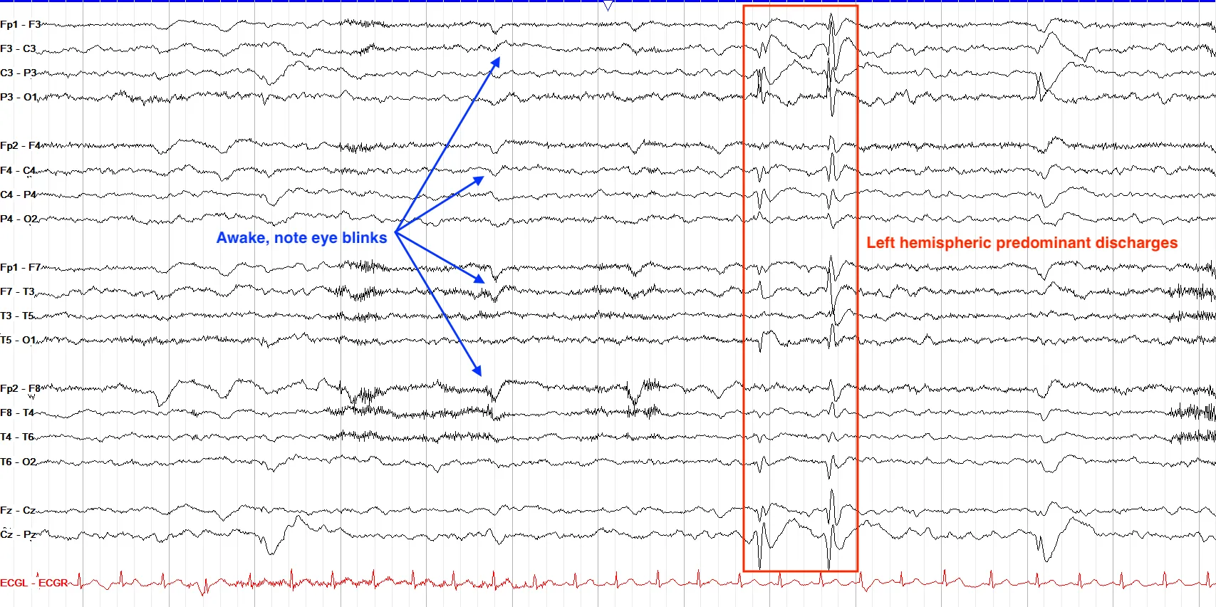
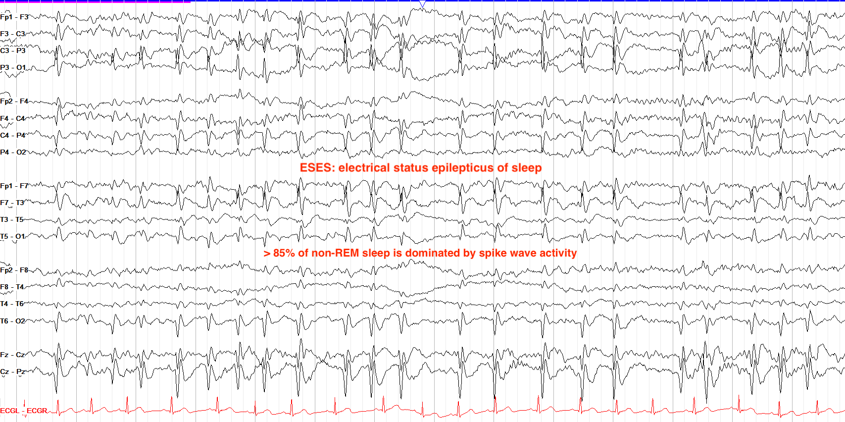
Electrical status epilepticus of sleep (ESES)--also called continuous spike and waves during slow wave sleep (CSWS)--describes a dramatic increase in epileptiform discharge burden during sleep, with discharges present in at least 85% of non REM sleep. ESES is commonly found in Landau Kleffner Syndrome, in which children have seizures and language regression after an initially normal development due to the presence of ESES on a nightly basis. In the example below, this patient has occasional left hemispheric predominant discharges when awake, which increase in frequency to essentially continuous during sleep.
Juvenile Myoclonic Epilepsy (JME)
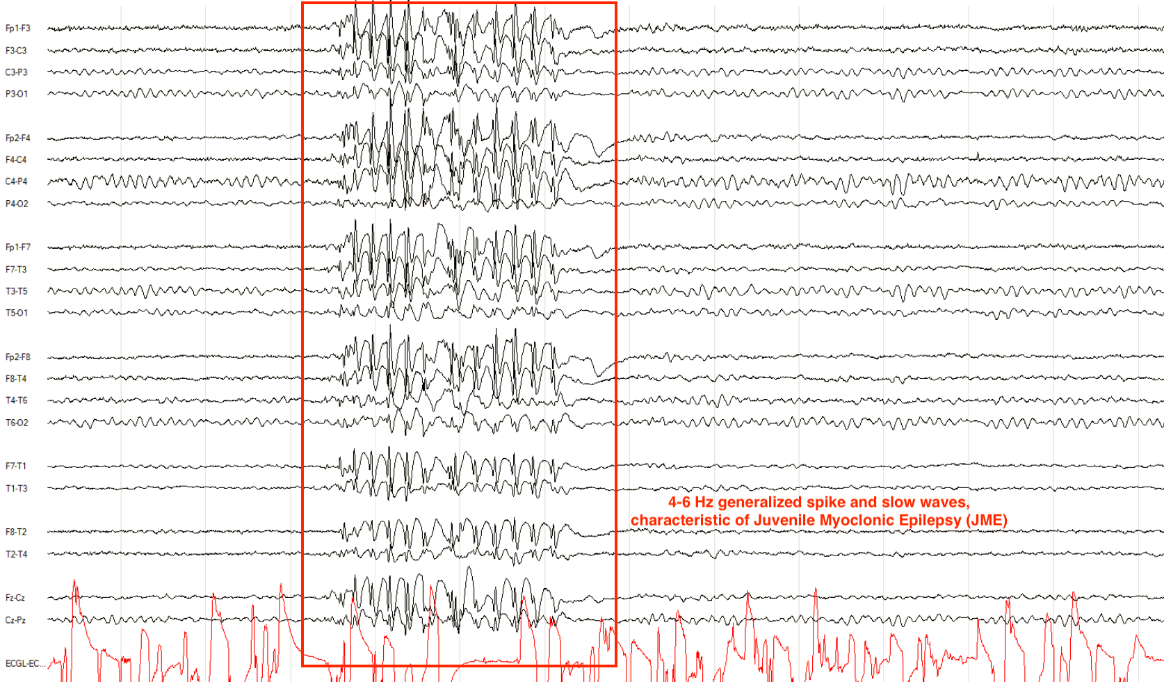
Juvenile myoclonic epilepsy is among the more common pediatric epilepsy syndromes, arising in adolescence with possible persistence into adulthood. Clinically it is marked by myoclonic jerks, more often in the morning, and on EEG you may see a classic, intermittent 4-6 Hz generalized spike or polyspike and slow wave pattern.
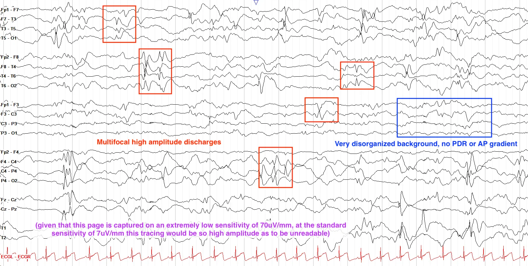
This page is captured at a sensitivity of 70uV/mm, which is very low for scalp EEG. So on the usual reading sensitivity of 7uV/mm, all the activity here would be so high amplitude as to be unreadable. Understanding that, this tracing shows a very high amplitude, disorganized background with multifocal epileptiform discharges, consistent with hypsarrhythmia.
- The pediatric EEG progresses from mostly delta, through theta, and finally to the alpha predominant adult tracing.
- The normal PDR progression is 4-5 by 6 mo, 6 by 1 yr, 7 by 2 yr, 8 by 3 yr, 9 by 8 yr, 10 by 10 yrs.
- Pediatric tracings tend to be higher amplitude with prominent sleep architecture including vertex runs and hypnagogic & hypnopompic hypersynchrony.
- Posterior slow waves of youth and slow/half alpha variants arise in the early years, and usually recede by late teens.
- Ohtahara syndrome is a devastating epileptic encephalopathy marked by discontinuity and high amplitude spikes.
- Hypsarrhythmia is a high amplitude, disorganized background with multifocal spikes, often seen with infantile spasms / west syndrome.
- Lennox Gastaut Syndrome (LGS) has a slow background with 1.5-2.5 Hz generalized spike waves.
- Self-limited epilepsy with centrotemporal spikes (SeLECTS, formerly BECTS) comes with nocturnal seizures, and usually resolves by adolescence.
- Absence epilepsy is marked by brief absence seizures with ~3 Hz generalized spike waves, provoked by hyperventilation.
- Electrical Status Epilepticus of Sleep (ESES) is marked by >85% discharge burden while asleep and causes development regression after initially normal development.
- Juvenile Myoclonic Epilepsy (JME) is classically associated with 4-6 Hz generalized spike wave activity.