Rhythmicity & Periodicity Run Along a Spectrum
We briefly discussed rhythmic and periodic patterns (RPP) in the terminology section; recall they are both classified as waveforms of similar morphology and duration that repeat six or more times contiguously. They differ in that periodic patterns are composed of discrete waveforms with gaps in between each, while rhythmic patterns are comprised of undulating waveforms that each flow directly into the next.
The clinical significance of RPP, however, is substantially more complicated than their basic definitions, as they can indicate issues from mild nonspecific dysfunction to markedly high seizure risk with status epilepticus. The stratification therein depends on a number of factors including the RPP’s distribution (generalized vs lateralized), frequency, persistence, morphology, and presence or absence of evolution. Patterns thus move along a spectrum of seizure risk, with the uncertain middle ground—where a pattern is neither clearly interictal nor ictal—termed the ictal-interictal continuum (IIC).
The IIC is a flexible framework that brings classification closer to the genuinely shifting nature of EEG, and understanding it is vital to accurate interpretation. The full criteria are in the ACNS Standardized Critical Care EEG Terminology 2021 Reference Chart.
Here we’ll discuss the varying types of RPP commonly encountered, in ascending order of seizure risk. Below is an interactive schematic that lets you walk through the IIC based on its contained patterns and modifiers per the 2021 ACNS criteria; this is not meant as a diagnostic guide or tool, but rather to help orient and guide familiarization with this fundamental aspect of EEG interpretation.
Interactive ictal-interictal continuum.
Generalized Rhythmic Delta Activity
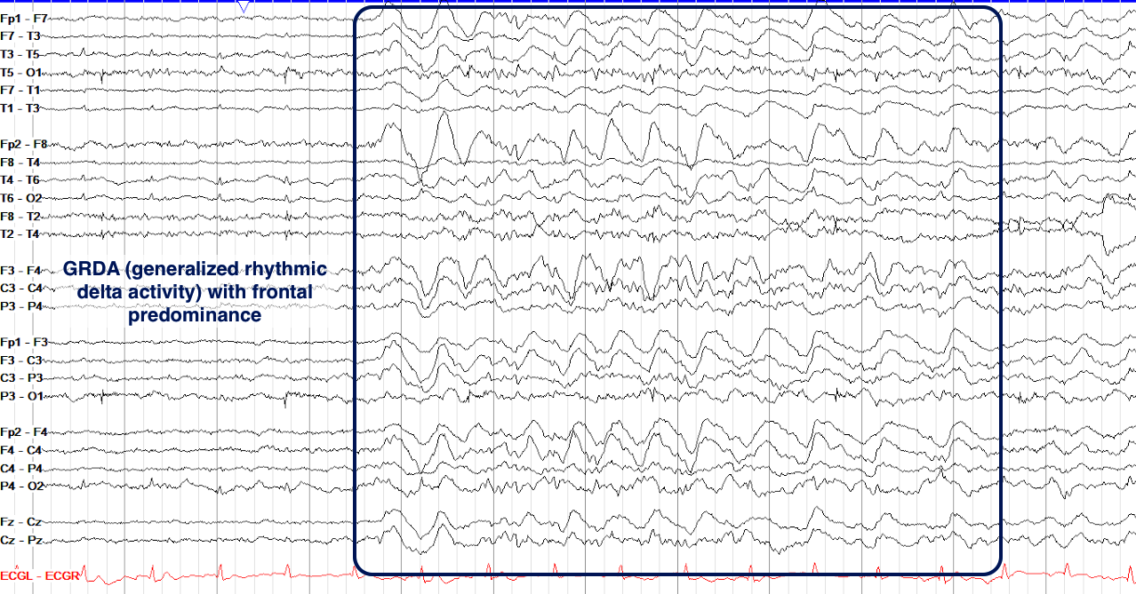
Among the most common abnormalities you’ll encounter in ill and hospitalized patients, generalized rhythmic delta activity (GRDA) sits on the nonspecific end of the spectrum in terms of etiology and significance. It is commonly seen in the setting of very ill patients with infections, metabolic derangements, and neurodegenerative disease (similar to generalized slowing as described above). GRDA can be quite diffuse but is often frontally predominant.
On the example below, note that the bifrontal delta activity does not travel back to the posterior regions; the current ACNS nomenclature terms this GRDA with frontal predominance, whereas older terminology would have overtly called it frontal intermittent rhythmic delta activity (FIRDA), and more truly generalized activity was previously called generalized intermittent rhythmic delta activity (GIRDA).
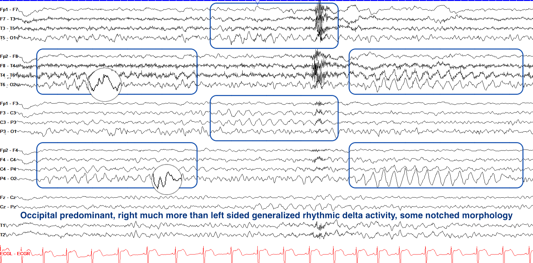
While frontally predominant GRDA is common with encephalopathy, occipitally predominant GRDA, previously termed occipital intermittent rhythmic delta activity (OIRDA), is more common in children with generalized epilepsy, though it is sometimes also found in those with nonspecific encephalopathy. In the example below note that the occipital rhythmic delta is much more prominent on the right side and has some notched morphology akin to blunted sharps; in cases such as this, the pattern likely leans toward being epileptogenic (more so if the notches are more overtly spikes or sharps).
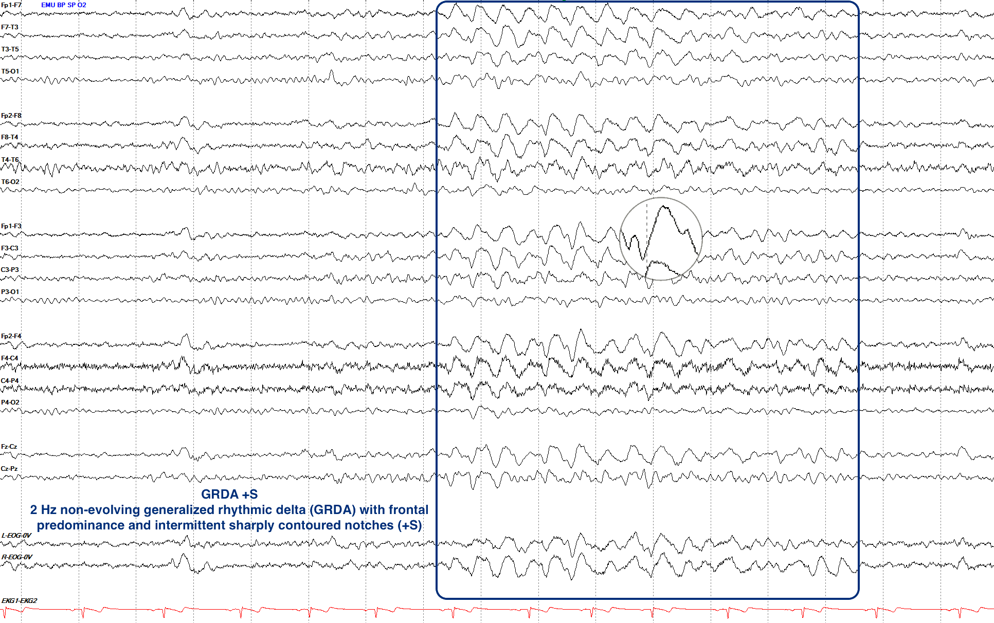
GRDA does not technically sit on the IIC, and under formal ACNS criteria is excluded from the continuum entirely as its seizure risk is far lower than the rest of the patterns we discuss here, even with plus features. That being said, if it acquires superimposed fast activity (+F), sharp activity (+S), or both together (+FS), in the right clinical context it can still suggest heightened seizure risk. Similarly, as with any of the RPP patterns, if GRDA starts to show clear evolution in its morphology, frequency or distribution, that becomes much more suspicious for seizure. Notice below how we have frontally predominant GRDA but within that rhythmicity exist sharply contoured notches in the frontal regions, too; this is an example of GRDA +S (sharp activity).
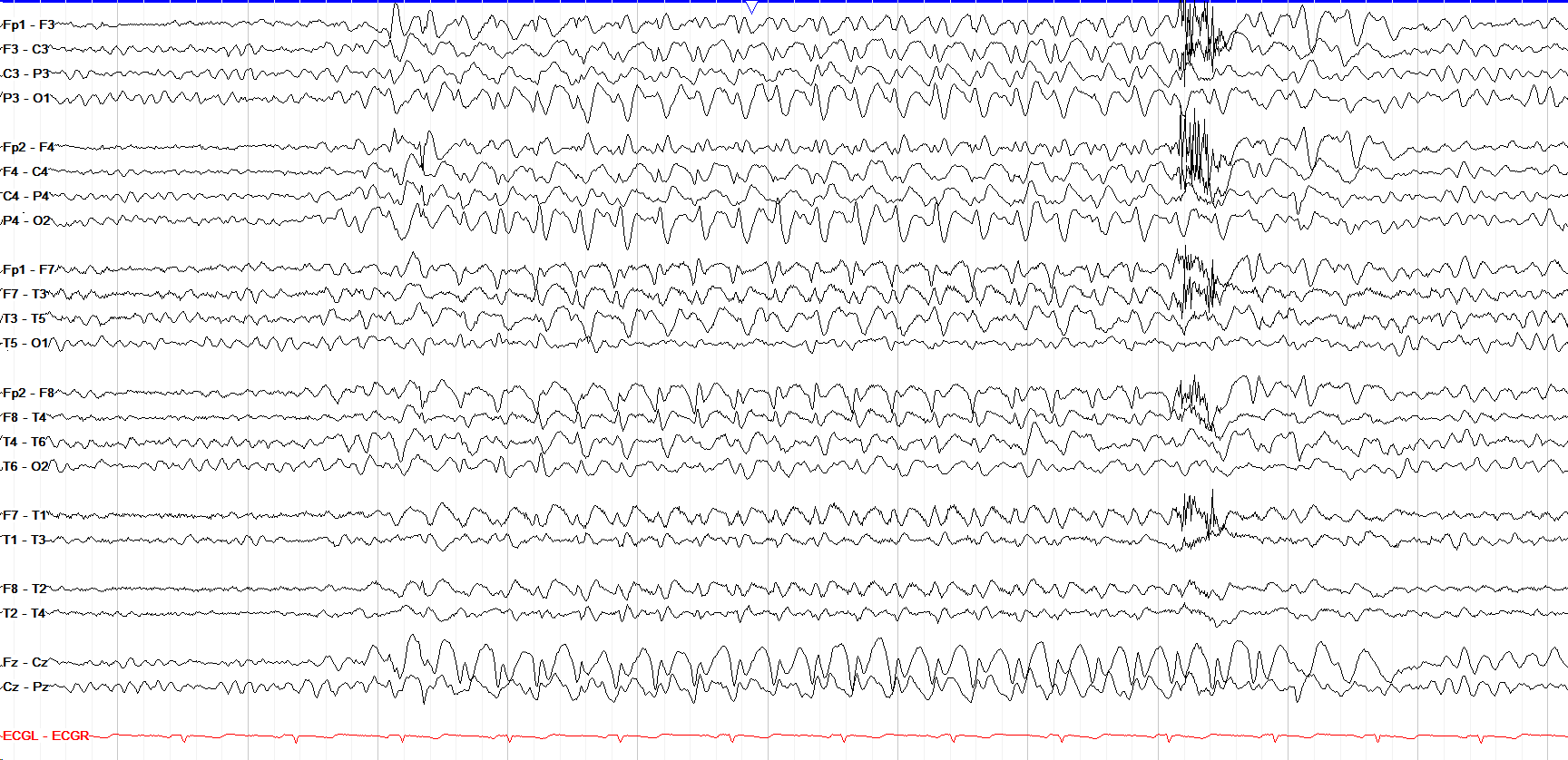
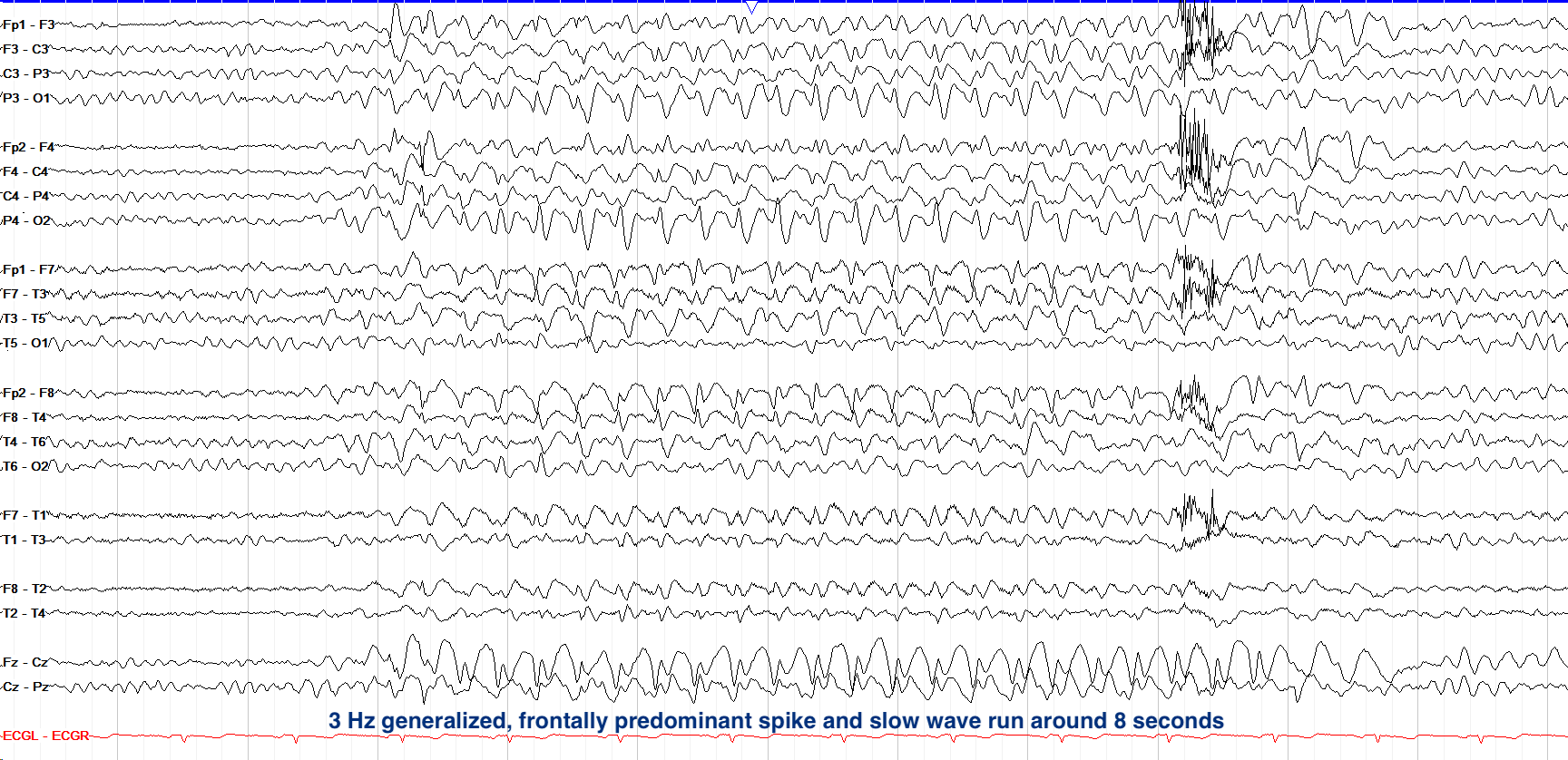
This image, while showing monomorphic activity, doesn’t actually show GRDA (or the other rhythmic/periodic patterns we’ve covered) but rather more overt spike-and-slow-wave activity that is fairly diffuse but frontally predominant (notice the well-formed spike-and-slow-wave morphology recurring regularly over the Fz region, especially). So rather than rhythmic delta with superimposed sharps (GRDA+S), this is a run of frank, overtly epileptiform discharges.
As such, it doesn’t sit in the low-risk, uncertain territory where GRDA+S sits; instead it’s at the upper ictal edge, because at 3 Hz this would meet electrographic-seizure criteria if it persisted for 10 or more seconds (at 8 seconds it just barely falls short). It shouldn’t be confused with a seizure (which requires 10 or more seconds, or a clinical correlate if briefer), nor with a BIRD, which can look similar (a brief, evolving ictal-appearing run) but requires a frequency >4 Hz. This tracing trends at only 3 Hz—a frequency classically seen with generalized epilepsy syndromes.
Generalized Periodic Discharges
Moving up the spectrum from GRDA and being in many ways its periodic counterpart, generalized periodic discharges (GPDs) are discrete and bilaterally synchronous discharges often seen with toxic and metabolic encephalopathies, anoxic brain injury, and Creutzfeldt-Jakob disease (CJD). Unlike GRDA, GPDs can meet formal IIC criteria when they trend >1–2.5 Hz, or at 0.5–1 Hz with a plus modifier or fluctuation, but they often still sit on the lower end; as with all patterns on the IIC, particular features of the GPDs affect the seizure risk associated with them.
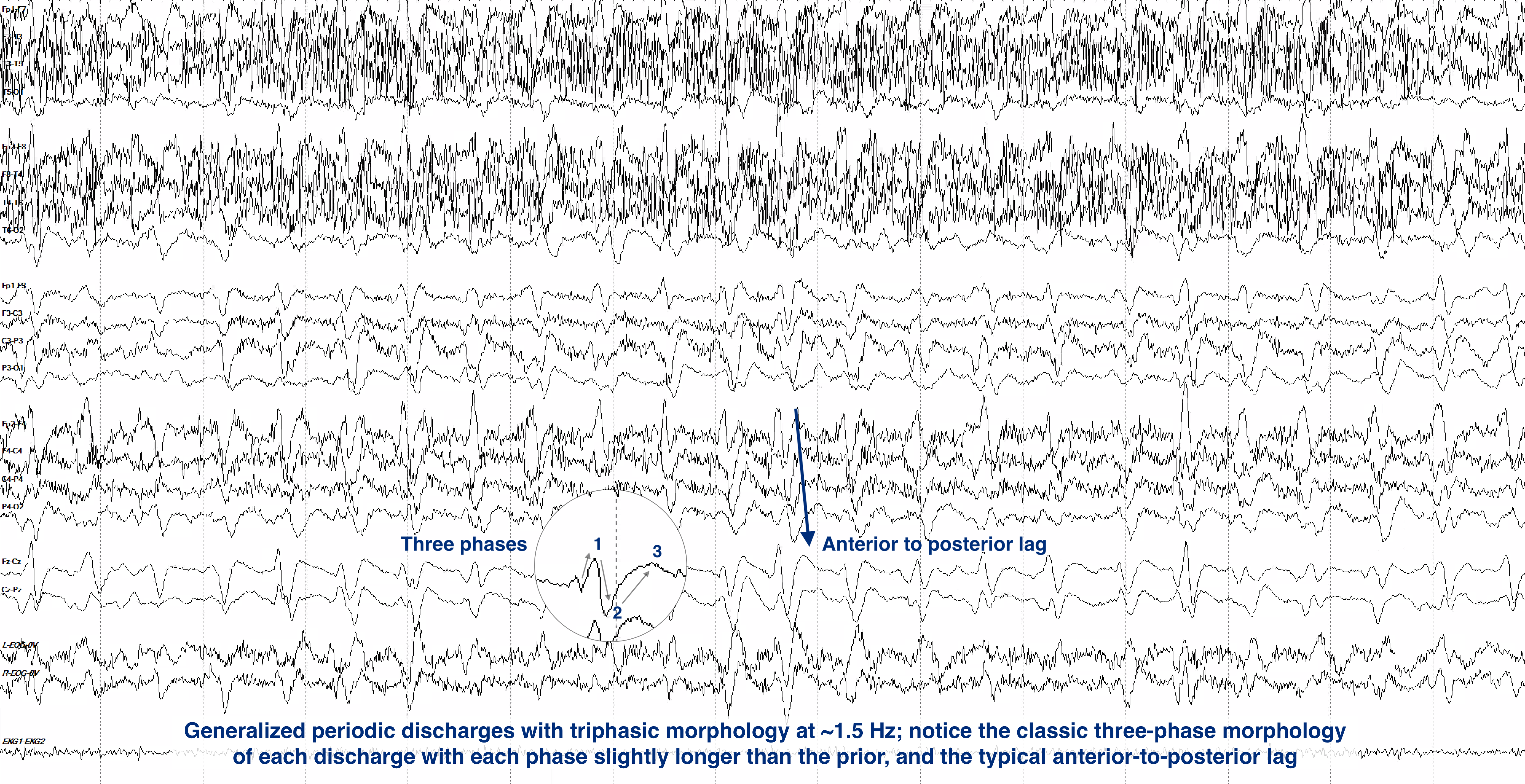
One of the more commonly seen GPD types are those with triphasic morphology (previously termed triphasic waves). Classically but not specifically associated with metabolic encephalopathy such as hepatic / renal failure or infection, the triphasic term is descriptive; these discharges are comprised of three phases, with each phase being slightly longer than the preceding one and with a subtle anterior to posterior delay in onset. In the example below we see a series of well-formed generalized discharges at around 1.5 Hz with that classic three phase morphology and anterior to posterior lag, though there is prominent overriding myogenic artifact throughout.
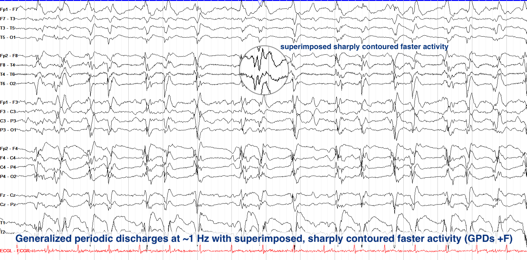
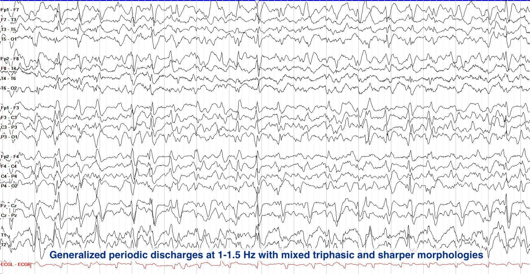
GPDs with frequency greater than 1.5–2 Hz or morphologies containing superimposed fast activity (+F) or rhythmic delta (+R) generally have higher seizure risk. Critically, if GPDs exceed 2.5 Hz for ten or more seconds, they are no longer considered interictal at all: they’ve shifted to an overt electrographic seizure (remember this cutoff—2.5 Hz for ten or more seconds—as that is the threshold for seizure for any periodic pattern). Below you can see GPDs at 1 Hz. At that frequency the ACNS criteria require a plus modifier or fluctuation for them to formally be on the IIC; in this example the superimposed sharply contoured faster activity (+F) does that. Note that the plus modifiers differ between rhythmic and periodic patterns; periodic discharges can have superimposed fast activity (+F), superimposed rhythmic delta (+R), or both (+FR), whereas rhythmic patterns can have superimposed sharp activity (+S), superimposed fast activity (+F), or both (+FS).
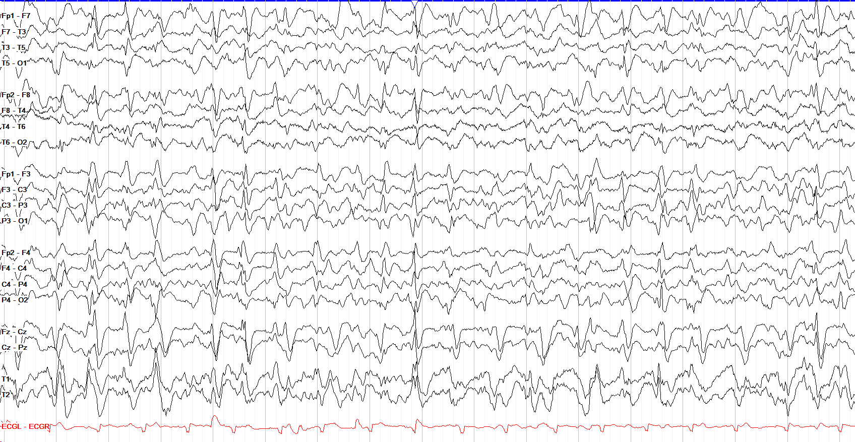
This tracing exemplifies the fact that all of the patterns along the IIC blur into one another; they truly are a continuum along both frequency and morphology, not discrete categories. While the discharges on this tracing show the classic three-phase and anterior-posterior lag morphology of the triphasic variant, they are also much sharper in appearance than standard triphasic forms. This kind of fluctuation and intermingling is common across studies—some patterns are quite textbook for a single entity while others can shift between sharpness, rhythmicity, periodicity and more.
Lateralized Rhythmic Delta Activity
When generalized, rhythmic delta is mostly nonspecific in origin and significance; lateralized rhythmic delta activity (LRDA), while sharing the same undulating delta but being seen in only one hemisphere or region, can meet formal IIC criteria when it trends >1 Hz with a plus modifier or fluctuation, and has a similar seizure association to periodic patterns. LRDA usually reflects a focal structural process in the rhythmic area or hemisphere, such as a stroke, tumor, or hemorrhage.
The tracing below shows prominent rhythmic delta over the right temporal region without a corresponding change over the left hemisphere; this is an example of temporal LRDA, which used to be called temporal intermittent rhythmic delta activity (TIRDA). Of note, temporal LRDA has a high association with mesial temporal lobe epilepsy.
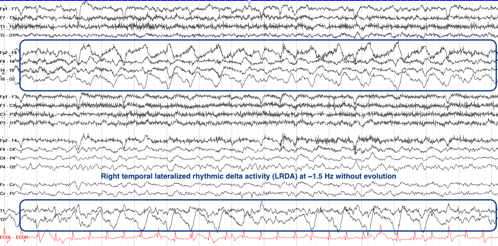
On the IIC, LRDA fluctuating around 1 Hz sits somewhere near the middle, but its risk for shifting to overt seizure increases with its frequency, particularly at 2 Hz or faster, while superimposed fast or sharp activity (+F/+S) and greater prevalence also push it higher on the IIC. Unlike with periodic patterns, rhythmic ones that reach 2.5 Hz do not implicitly shift into seizure territory, though any evolution still suggests true seizure activity.
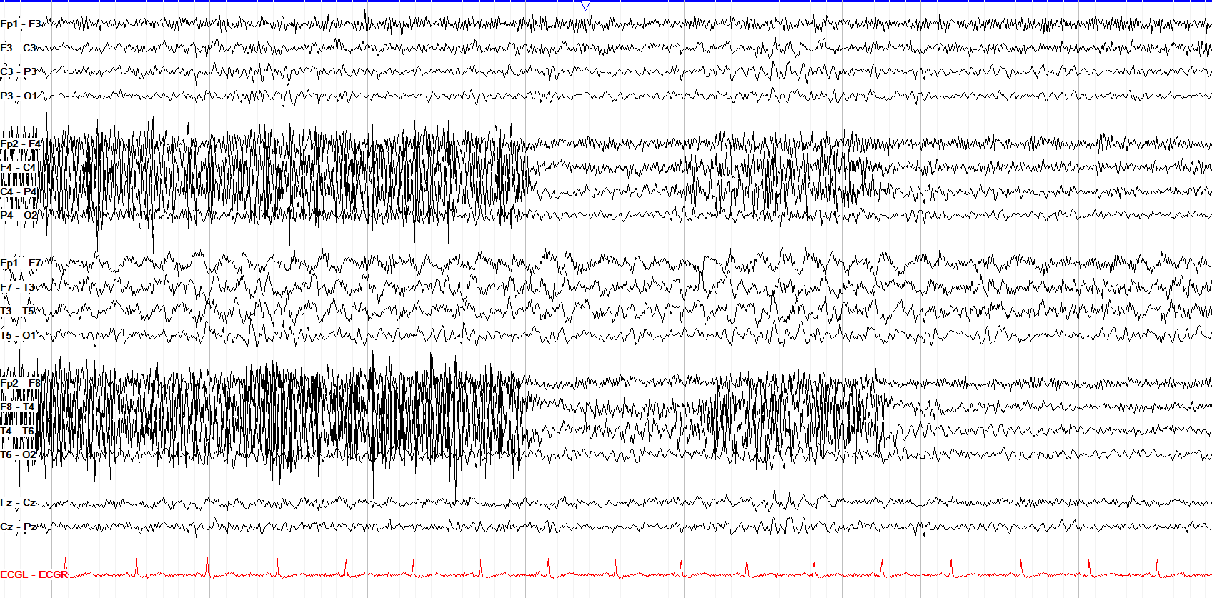
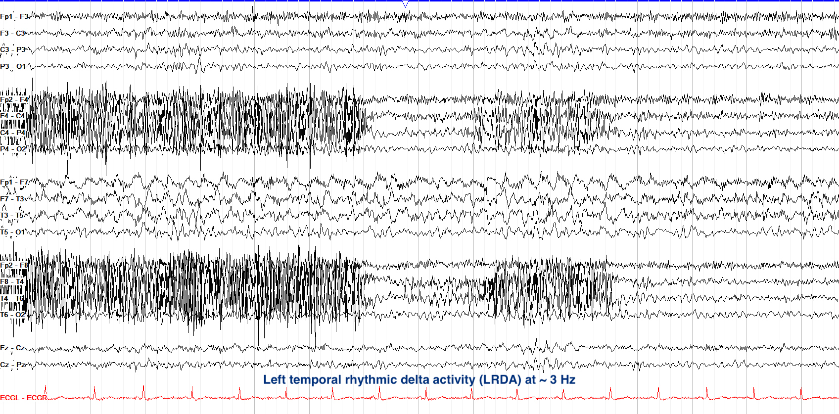
Here we see regular 3 Hz rhythmic delta activity over the left temporal region, though prominent myogenic artifact overlies it and could, at first glance, make you mistake the RDA for more polymorphic slowing (note the even heavier myogenic artifact largely obscuring the right side). Historically, temporal LRDA, previously termed TIRDA, has been most closely associated with mesial temporal lobe epilepsy.
This LRDA lasts ten or more seconds, but remember that while a periodic pattern exceeding 2.5 Hz for ten or more seconds shifts into overtly ictal territory, rhythmic delta activity doesn’t follow that frequency rule but rather remains on the ictal-interictal continuum up to 4 Hz as long as it doesn’t evolve. Superimposed sharp or fast activity would raise its risk on the IIC, but doesn’t inherently make it ictal.
Lateralized Periodic Discharges
In the upper half of the IIC sit lateralized periodic discharges (LPDs) over one hemisphere or region. LPDs carry the strongest baseline seizure association of the standard rhythmic or periodic patterns and are very often tied to acute focal pathology such as stroke, tumor, abscess, and (especially in the temporal regions) herpes simplex encephalitis. On the example below, note the discrete periodic discharges recurring roughly once per second, lateralized to the right hemisphere with a clear interval between each. Older terminology called these PLEDs (periodic lateralized epileptiform discharges).
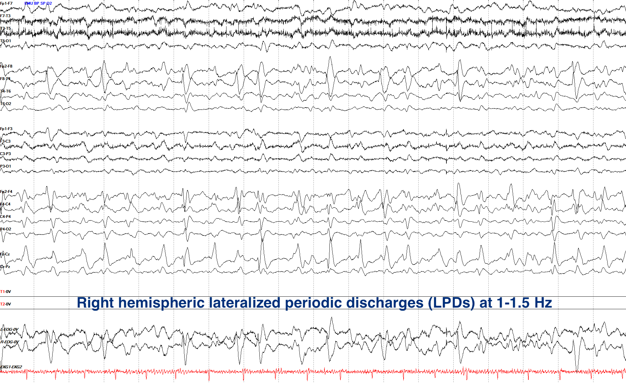
LPDs generally sit high on the IIC at baseline; very slow ones (less than 1 Hz) have much less seizure risk, though, while the usual cohort of faster frequency, superimposed fast and / or rhythmic activity (+F/+R or +FR), and greater prevalence increase the risk for seizures with LPDs even further. When two independent foci discharge out of phase with one another, the pattern is bilateral independent periodic discharges (BIPDs, formerly BIPLEDs), and this pattern can suggest a seizure risk even higher than unilateral LPDs.
As with other periodic discharges, LPDs faster than 2.5 Hz for ten or more seconds, or with clear evolution, are considered overt electrographic seizures.
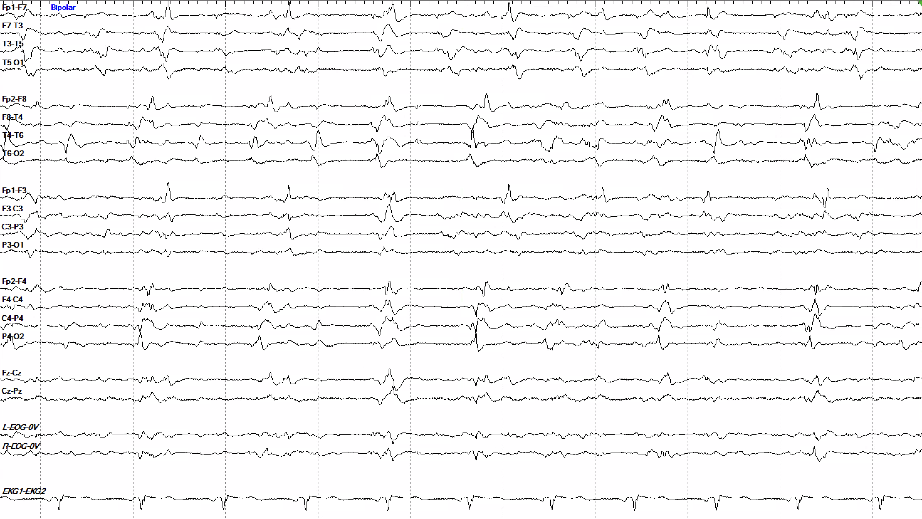
Here we see what look like two separate groups of 1 Hz discharges on the left and right sides. This could be consistent with either asynchronous GPDs or bilateral independent periodic discharges (BIPDs), though it can be difficult to tell them apart from a single page. When you’re reading a real tracing, one way to differentiate the two possibilities is by looking at the broader record; if the tracing overall shows an asynchrony with the left and right sides being slightly out of sync with each other, asynchronous GPDs are more likely, but if the tracing is largely synchronous except for the discharges then BIPDs are more likely. It’s important to clarify which pattern this actually is because that determines where they fall on the IIC; asynchronous GPDs at ~1 Hz sit on the lower end, whereas BIPDs sit higher.
Brief Potentially Ictal Rhythmic Discharges
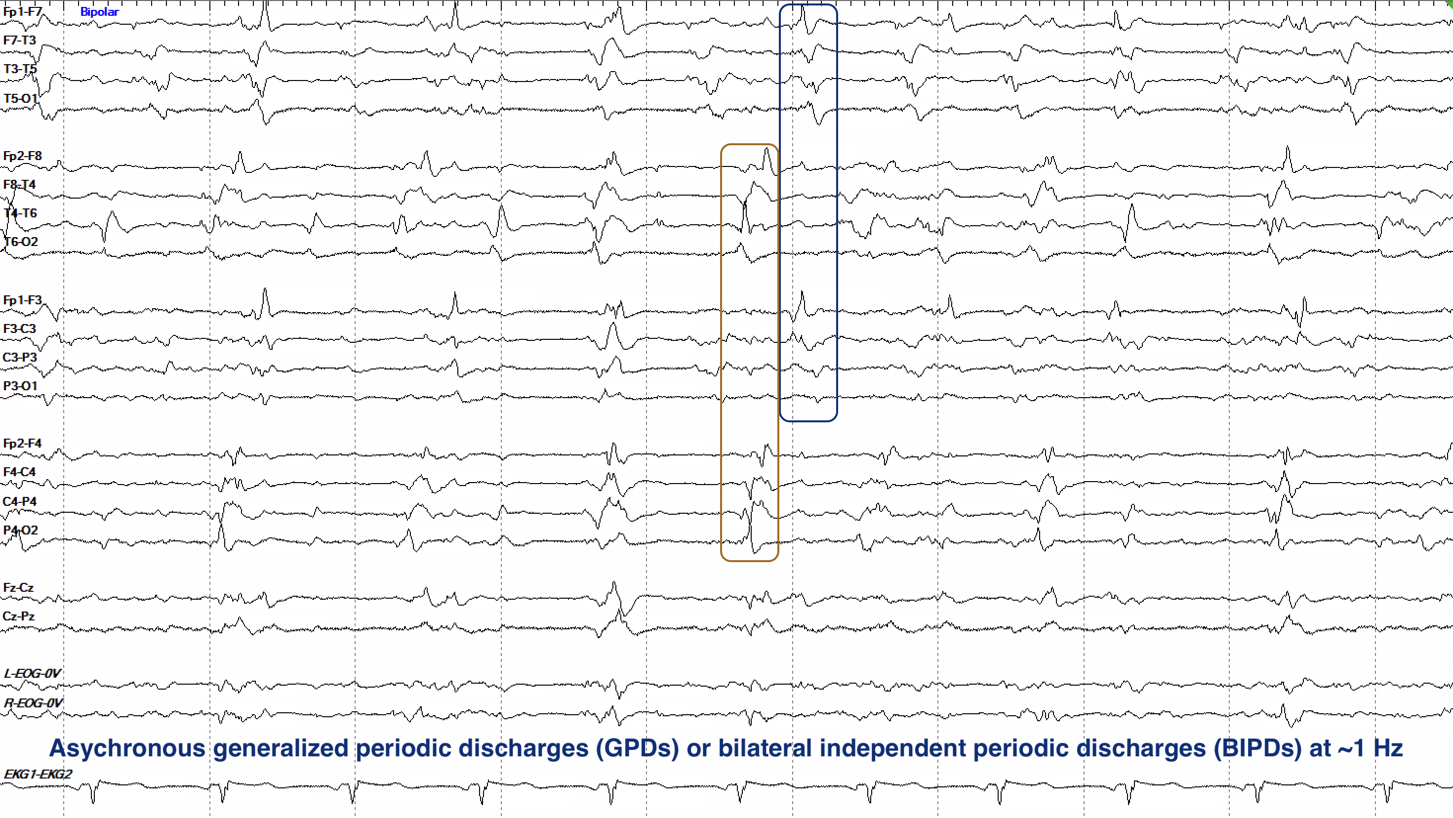
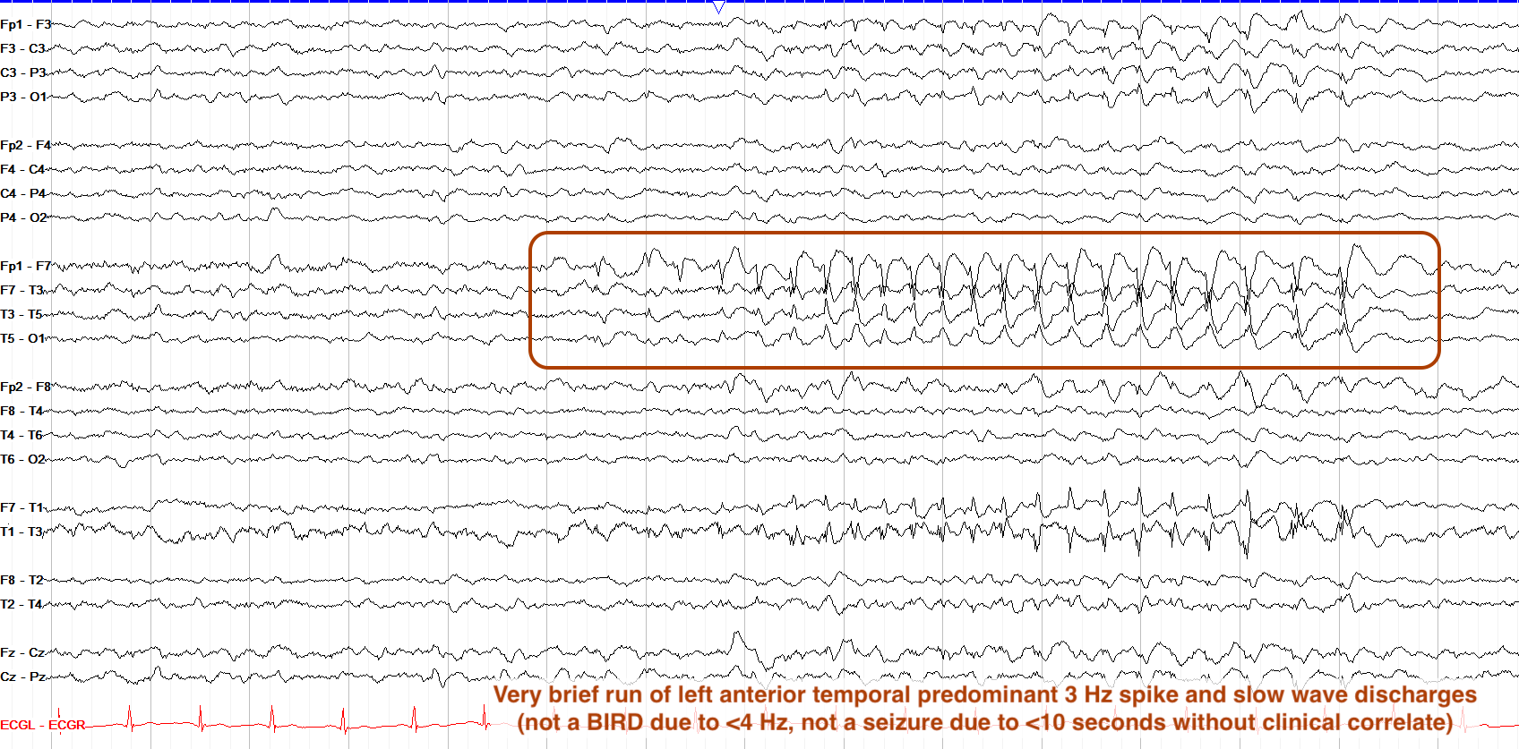
Sharing a name but little else with their winged brethren, brief potentially ictal rhythmic discharges (BIRDs) are ictal-appearing rhythms of greater than 4 Hz that do not meet the minimum 10-second criteria to be considered a seizure. Those that clearly evolve or resemble a patient’s own interictal discharges or seizures are termed definite BIRDs, while those that don’t, and are merely sharply contoured, are termed possible BIRDs. Definite BIRDs in particular sit closest to true seizures on the IIC, as the only real difference is their duration; they show ictal evolution, morphologies, frequencies and more, so a BIRD that exceeds its 10-second limit is by definition a seizure. BIRDs are hewn so closely to seizures, in fact, that well over half of tracings with BIRDs will eventually show true electrographic seizure activity.
In the example below, we see a 7-second run of largely non-evolving right temporal spike-wave activity consistent with BIRDs; note the field into the right frontal region.
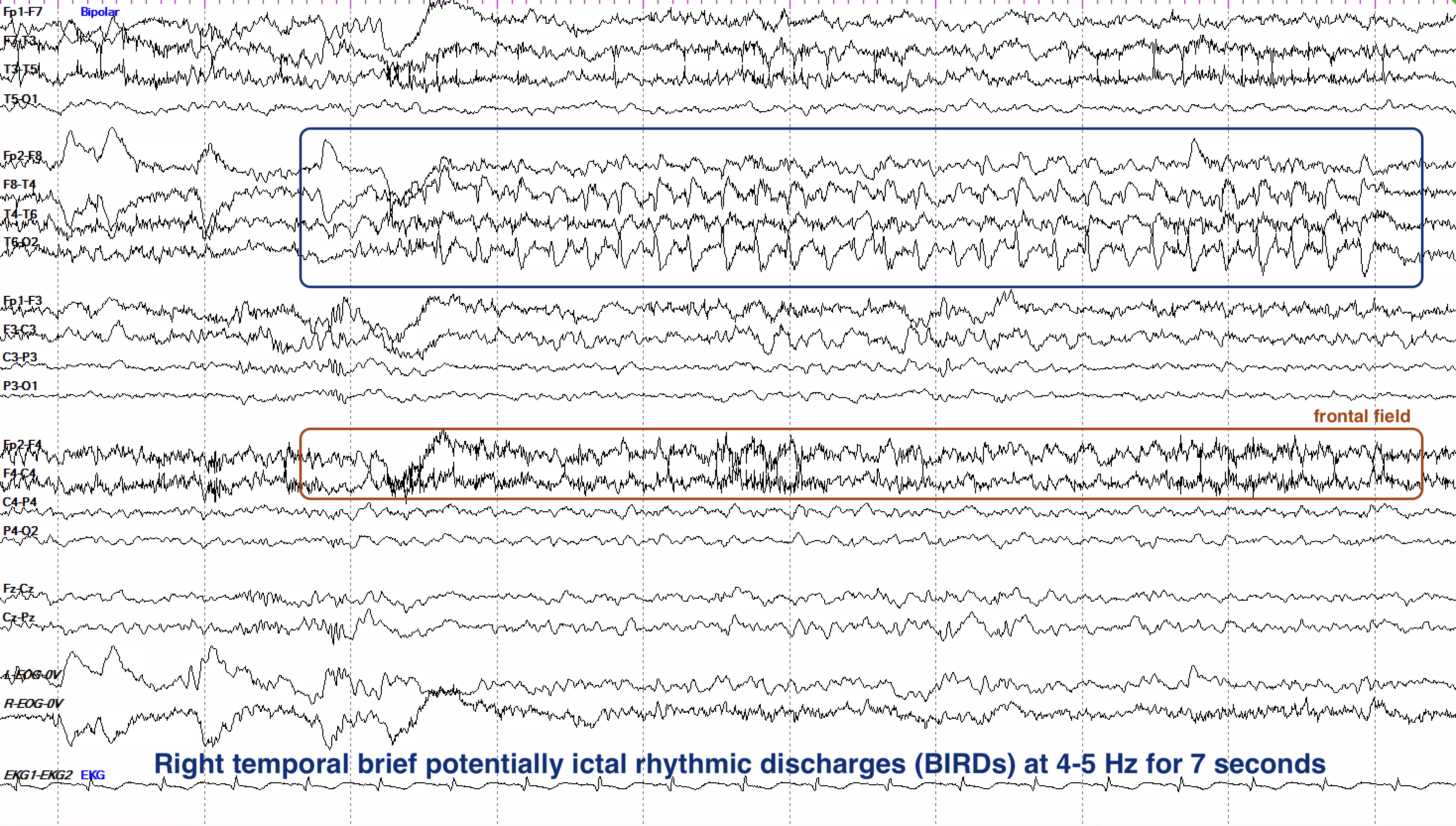
BIRDs can be seen in a broad range of patients, but most commonly in those with critical illness, newfound tumors and strokes, neonates, and certain autoimmune encephalitides such as NMDA receptor encephalitis.
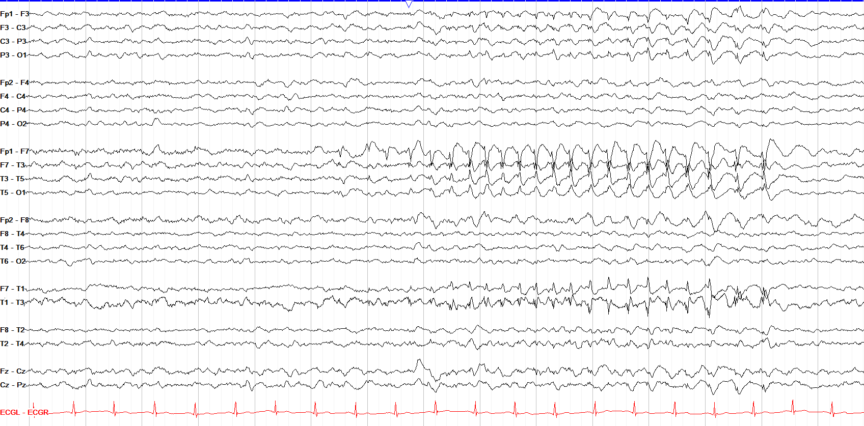
Here we see a background of low-to-moderate amplitude delta and theta frequencies with a clear 7–8 second run of left anterior temporally predominant spike-and-slow-wave activity, with field into the left frontocentral region. That activity builds in amplitude and organization before slowing slightly to offset (so it shows some evolution), which might make you lean toward calling it a BIRD or seizure. However, BIRDs formally require more than 4 Hz and this is only 3 Hz, and while periodic patterns can become seizures if they exceed 2.5 Hz for ten or more seconds, this one is shorter than that and thus not a seizure, either. So overall it’s best described as a brief ~3 Hz run of left anterior temporal spike-and-wave discharges.
SIRPIDs
Stimulus-induced rhythmic, periodic, or ictal discharges (SIRPIDs) are another aptly named entity. They aren’t one specific waveform or pattern, but rather a description for when any of the patterns we’ve discussed here (GPDs, GRDA, LPDs, LRDA or even a seizure) arises due to stimulation. SIRPIDs are often seen in critically ill patients, and the stimulus for them may be subtle and easily missed on video, so it’s important to look closely; a noise, touch, or routine bedside care may be the trigger. Note that normal reactivity generally doesn’t show the kind of distinct rhythmic or periodic morphology of SIRPIDs.
Despite their name, SIRPIDs are not inherently ictal, though in terms of the IIC all the usual factors that push their underlying pattern into ictal territory still apply, including frequency, morphology, evolution, plus modifiers, duration, and clinical correlate.
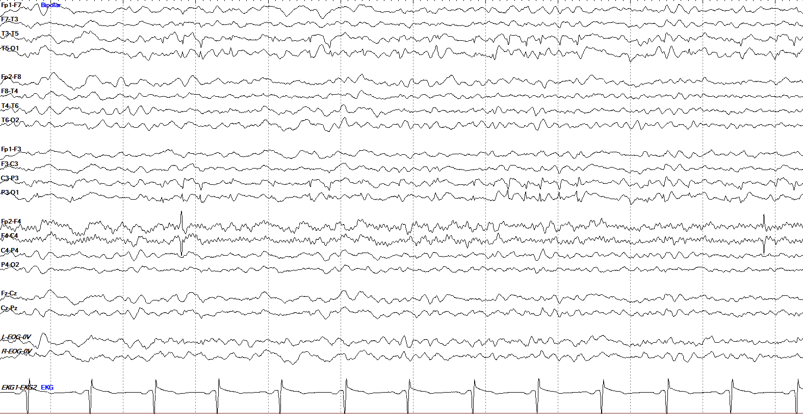
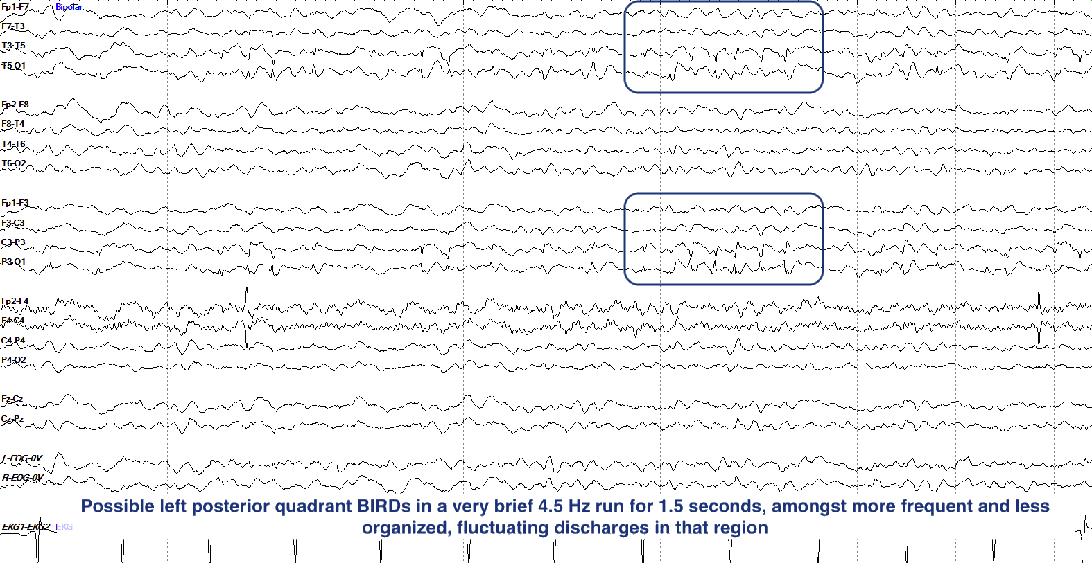
Yes, this pattern sits on the IIC, and close to the ictal end. Among the mostly delta and theta background of this tracing we see left temporo-parietal (T5 and P3 max) sharp waves, some with sharp and slow wave morphology that are initially poorly formed, quasi-periodic, and fluctuating. In the marked box, however, they don’t fully evolve but do build up into a more organized 4.5 Hz run for about a second and a half, so meet the criteria to be considered BIRDs (>4 Hz for at least 6 cycles). What makes these possible rather than definite BIRDs is that, formally, ACNS reserves “definite” BIRDs for those that clearly evolve or match one’s own interictal discharges or seizures.
- Rhythmic and periodic patterns (RPP) are monomorphic groups of waveforms that repeat six or more times, with periodic patterns having discrete discharges with gaps in between, while rhythmic patterns flow continuously.
- The significance of RPP ranges from nonspecific dysfunction to high seizure risk, based on their position on the ictal-interictal continuum (IIC), an uncertain middle ground between clearly interictal and ictal patterns that reflects seizure risk; the IIC is graded by pattern type, distribution, frequency, prevalence, morphology, plus-modifiers, and evolution.
- GRDA is nonspecific and formally excluded from the IIC, even with plus features. However, +F or +S still raise concern without moving it onto the continuum, while clear evolution makes it a seizure.
- GPDs sit on the IIC, with +F, +R and faster frequencies marking higher risk.
- LRDA has a seizure association similar to periodic patterns on the IIC, but its route to definite seizure is via evolution rather than frequency alone.
- LPDs carry the strongest baseline seizure association and are tied to acute focal pathology; BIPDs (two independent foci) are even more concerning.
- Any periodic pattern faster than 2.5 Hz for at least 10 seconds, or with clear evolution, is an electrographic seizure (this frequency threshold does not apply to rhythmic delta).
- BIRDs are >4 Hz ictal-appearing discharges under 10 seconds; those that evolve or resemble one’s own interictal discharges or seizures are definite BIRDs, while merely sharply contoured runs are possible BIRDs. They sit closest to the ictal end of the IIC, and most such tracings go on to show electrographic seizures.
- SIRPIDs are stimulus-induced RPPs or seizures, and can appear as any of the other patterns as they refer not to a specific pattern, but rather the stimulus trigger for a pattern.