Seizures Should Evolve
A seizure is an abnormal, organized and evolving burst of cortical activity that interrupts the brain's usual function. Clinically, they can present as anything the brain can experience, including sensorimotor activity, emotion, autonomic changes and more.
Electrographically, like interictal activity, seizures (aka ictal activity) can be either generalized or focal but either way seizures must evolve in frequency, morphology or distribution, and they should last more than ten seconds unless there is a clear clinical correlate, in which case they can be briefer. Interictal activity can fluctuate and become very frequent, but fluctuation is not equivalent to evolution, and if it never evolves in waveform shape, changes in frequency of those waveforms, or spreads in its distribution, it is largely not considered a seizure.
There are exceptions to this requirement for evolution but they’re specific; namely, periodic patterns that exceed 2.5 Hz for ten or more seconds (previously discussed in the context of the IIC), absence seizures whose discharges arise and abate without much evolution, and myoclonic or other seizures that have discharges with a clear, time-locked clinical correlate. Generally speaking, though, seizures require evolution.
Generalized Seizures
Generalized seizures are marked by a non-localizable onset, often involving most or all areas but sometimes having anterior or other regional predominance. As with nearly any seizure type, the exact appearance varies widely, but must evolve in frequency (i.e. become faster or slower until offset), morphology (i.e. changing shape in the waveforms as it goes), and / or distribution (i.e. spread from one region to another).
Generalized seizures are most often seen with generalized epilepsy syndromes; subtypes of generalized seizures are discussed below and include generalized tonic clonic (GTCs, previously termed grand mal), absence, tonic, myoclonic, and atonic seizures. Any time you see a generalized seizure, look closely to ensure that it isn't actually a focal to bilateral tonic clonic seizure, in which the onset is localized but there is rapid propagation into both hemispheres.
On the example below, note that it lasts about twelve seconds and evolves across time, starting with sudden onset generalized rhythmic theta activity for about a half second, then high amplitude spike wave activity at ~3 Hz, which slows to about 2 Hz and then slows further to 1.5 Hz before offset towards the end of the page.
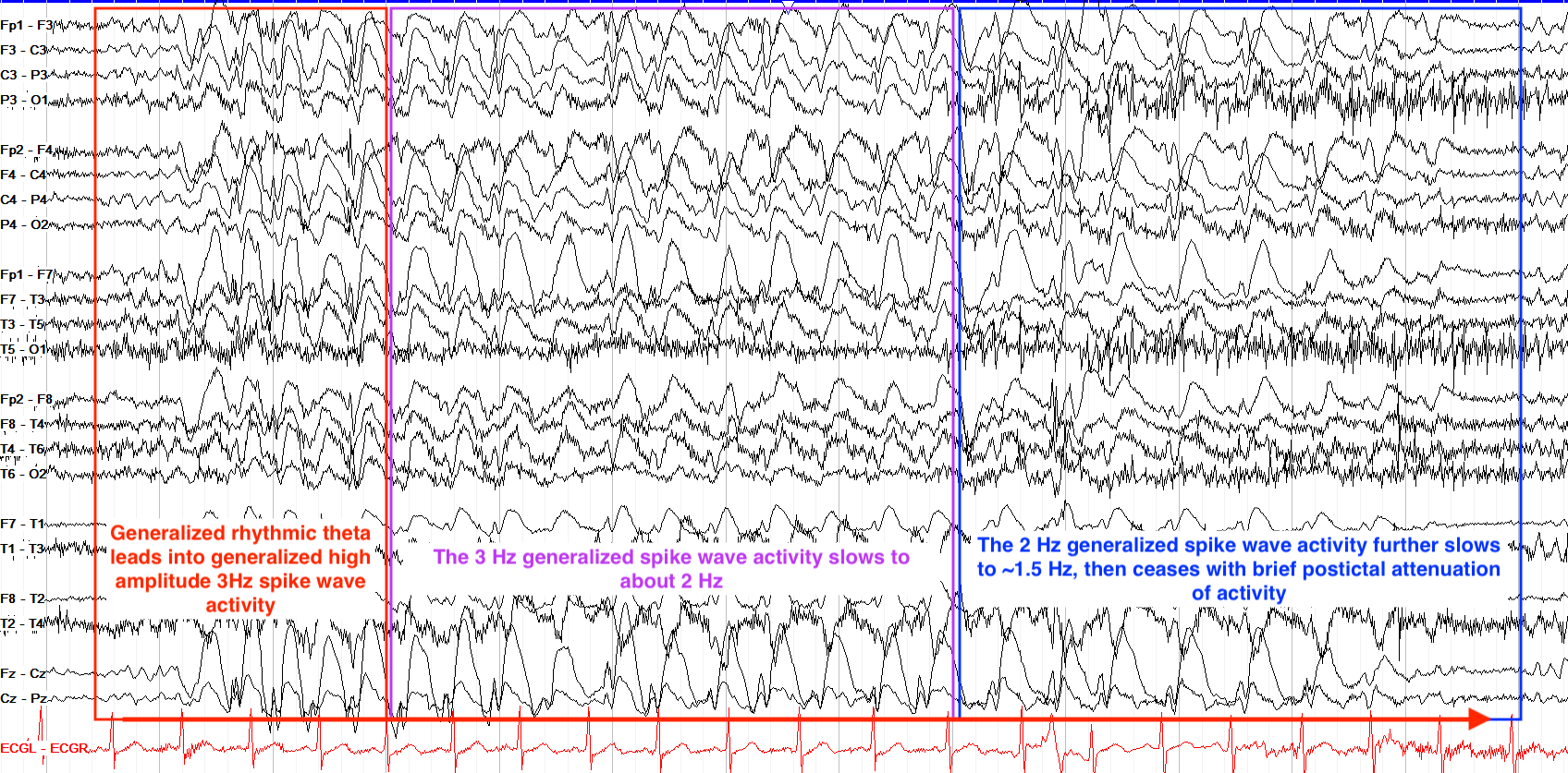
Most generalized seizures lead to impaired awareness or overt loss of consciousness because consciousness requires at least one cerebral hemisphere to be active along with the brainstem reticular activating system, and generalized seizures cause bilateral hemispheric dysfunction even if only momentarily. The main exception to this is myoclonic seizures, which are so brief that consciousness and awareness are typically preserved. This is particularly important for generalized non-convulsive seizures, which have minimal or sometimes no apparent outward evidence of seizures even as there may be internal interruption of consciousness or awareness, leading to lost time, memory impairment, attentional issues, and cognitive decline (note that non-convulsive seizures, sometimes termed subclinical seizures, can be either focal or generalized).
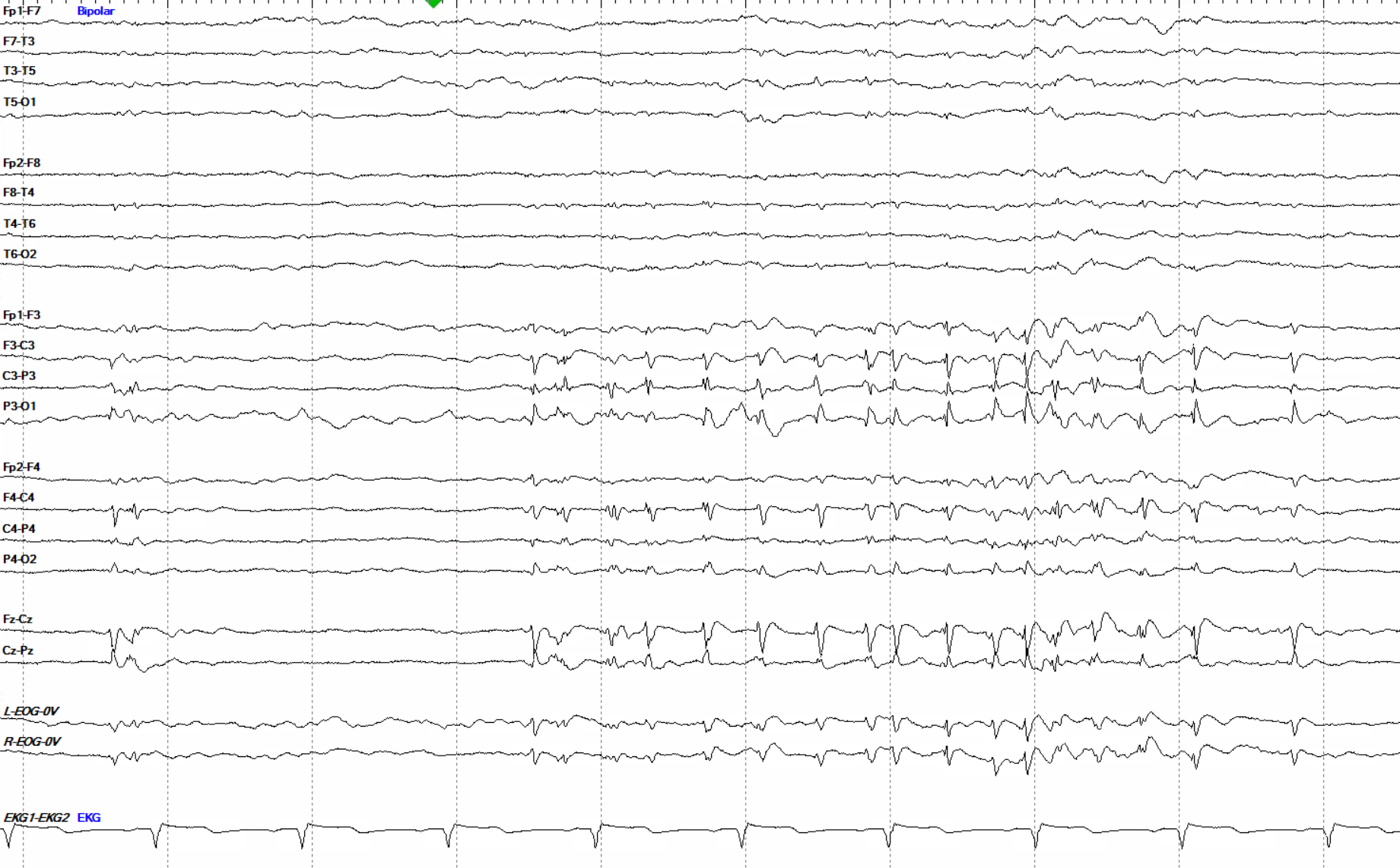
Here we see a diffuse low amplitude background against which an approximately 5.5 second burst of 2.5 to 3 Hz sharps emerge from the left more than right central predominant region. Across their span, they stay pretty much the same shape, do not increase or decrease in frequency, and don’t spread into other regions of the brain. So overall they don’t show any of the three key areas of evolution that most seizures require. Similarly, while they do trend at 2.5 Hz - 3 Hz, which passes the threshold for periodic patterns to shift into seizure activity on the IIC, they don’t persist for more than ten seconds. So they are not considered a seizure by either standard.
Focal Seizures
Focal seizures originate from a single area. They can be categorized into focal preserved consciousness, focal impaired consciousness, or focal to bilateral tonic clonic seizures (formerly focal aware, focal impaired awareness, and secondarily generalized; the oldest terminology used simple partial and complex partial for the first two).
In focal preserved consciousness (formerly focal aware) seizures one retains consciousness throughout the seizure, so could, for instance, continue talking and interacting while their left arm twitches or tremors with seizure activity. Because of this retained consciousness, they can remember the full extent of the seizure from beginning to end. Focal impaired consciousness (formerly focal impaired awareness) seizures, on the other hand, cause one to lose awareness and thus not recall the seizure, and often the time preceding and following the seizure, afterward. Both types of focal seizures can lead to isolated or mixed sensory, motor, emotional, autonomic or other symptoms.
If either type of focal seizure spreads to the contralateral hemisphere with tonic-clonic semiology (semiology being the physical manifestation(s) of the seizure), it becomes a focal to bilateral tonic clonic seizure, and looks clinically like a generalized tonic clonic seizure from that point onward. It is important, for such seizures, to pay attention to the early clinical signs and electrographically trace them back to the beginning to find their focus, and not just assume they are generalized from the start.
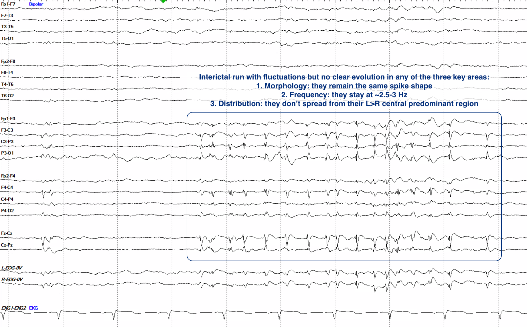
In the example below, note the onset of initially poorly formed delta activity over the left anterior temporal region that first evolves into more overt spike and slow waves maximal over F7. Within a second or two those discharges increase in amplitude and become much more well-formed at 2.5-3 Hz with a subtle field into the left central region; that central field becomes more prominent as the left anterior temporal discharges continue to increase in amplitude and then subtly slow prior to offset, with a few lingering left anterior temporal discharges that then stop; there remains quasirhythmic delta in the left temporal region that is baseline for the tracing overall. Note that this seizure never spreads into the right hemisphere.
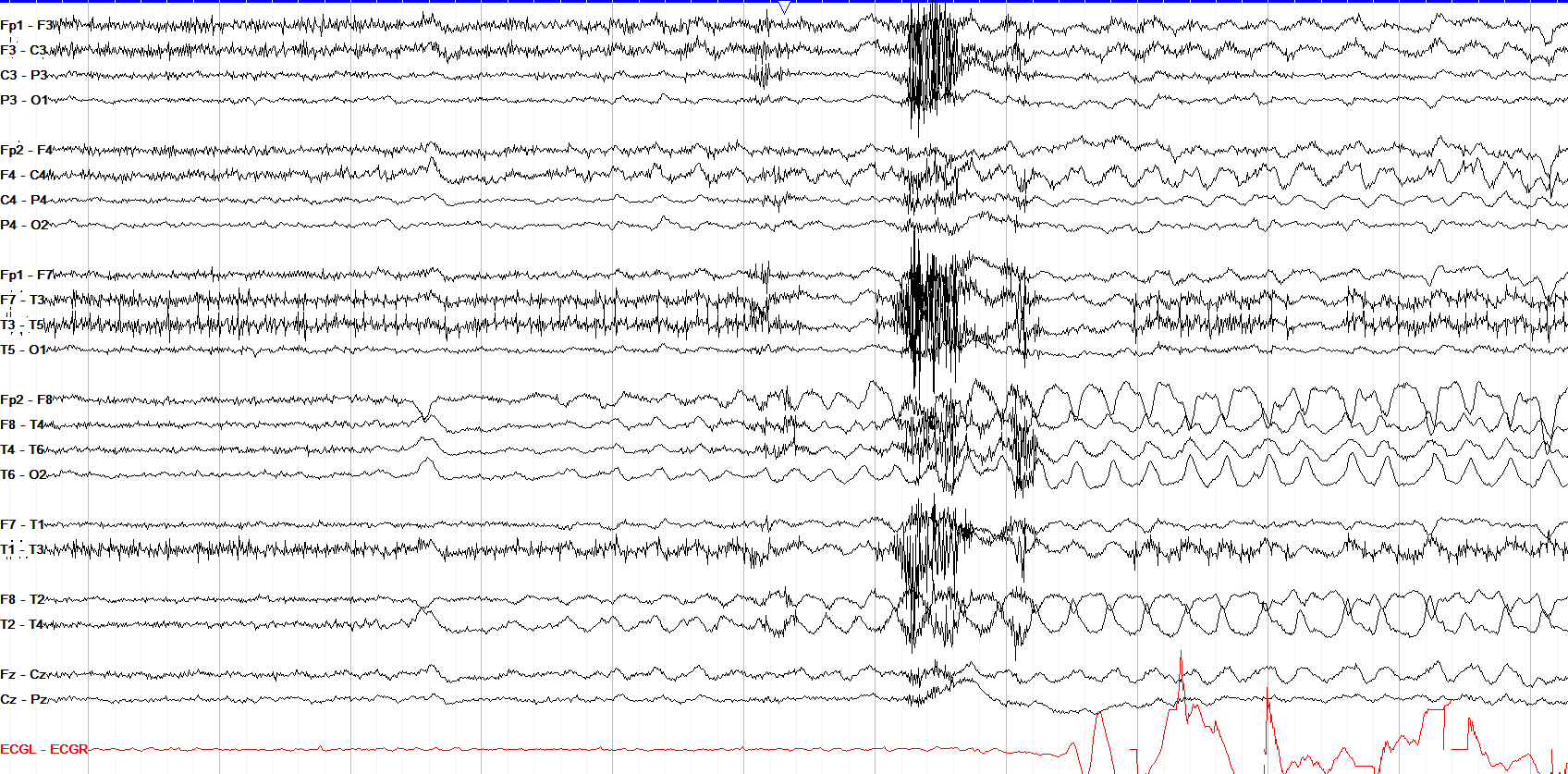
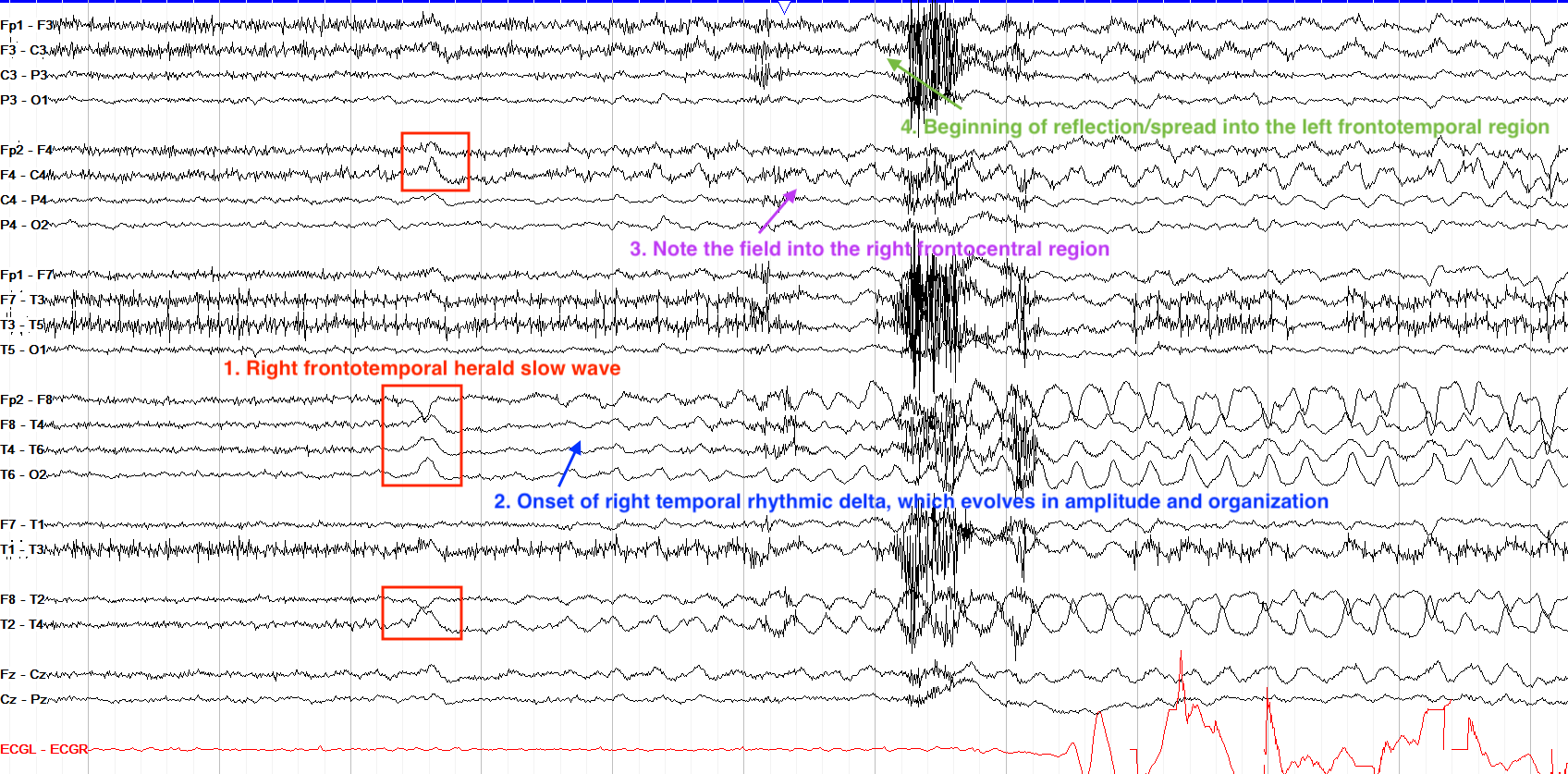
We only see the beginning of this seizure so we don’t know how it progresses, but it appears to begin with a very blunted / soft herald slow wave in the right frontotemporal region, which leads into subtle rhythmic delta of the right temporal region that increases in amplitude, becomes more organized at ~3 Hz, and spreads more broadly throughout the right frontotemporal region. There is some field into the left frontotemporal region, but in this single page we can’t tell if this evolves further into a fully bilateral seizure.
Tonic Clonic Seizures
Tonic clonic seizures are the prototypical seizure, previously termed "grand mal." They can be either generalized tonic clonic (GTC) or focal evolving to bilateral tonic clonic. They are marked, as their name suggests, by an initial period of continuous, tonic motor activity that clinically appears as stiffening and on EEG appears as significant and usually diffuse myogenic artifact. With such intensive depolarization, though, inhibitory processes come online to try to stop it, so that depolarization becomes less continuous and the tracing evolves into intermittent bursts of spike / polyspike and slow waves, with each spike burst corresponding to the clonic jerks that you see clinically.
Typically, after such intense neuron activity, the seizure is followed by a period of postictal attenuation and/or slowing, reflective of depleted ATP energy stores from the seizure’s massive metabolic demand, before an eventual return to the normal background. While seizures can be tonic clonic, they can also be solely tonic or solely clonic.
On the generalized tonic clonic seizure example below (the sensitivity of which is set at 20uV/mm to better show the high amplitude activity) note the progression from the tonic into clonic phases toward the end of the first page and across the second page, and how this seizure evolves in terms of frequency and morphology. Note also the period of postictal attenuation before the return of the background. While this particular example is generalized, if the onset was focal but there was evolution into both hemispheres, that would be termed focal to bilateral tonic clonic.
This seizure may at first appear generalized in onset, but if you look closely you can see a left centrotemporal sharp (C3 / T3 max) of moderate amplitude with a broad left frontocentrotemporal field immediately prior to the onset of a diffuse slow wave with superimposed generalized fast activity leading into sharply contoured theta into delta activity that progresses to very high amplitude generalized, frontally predominant spike wave activity at 1-1.5 Hz, which on the second page slows to 0.5 Hz prior to offset.
Tonic Seizures
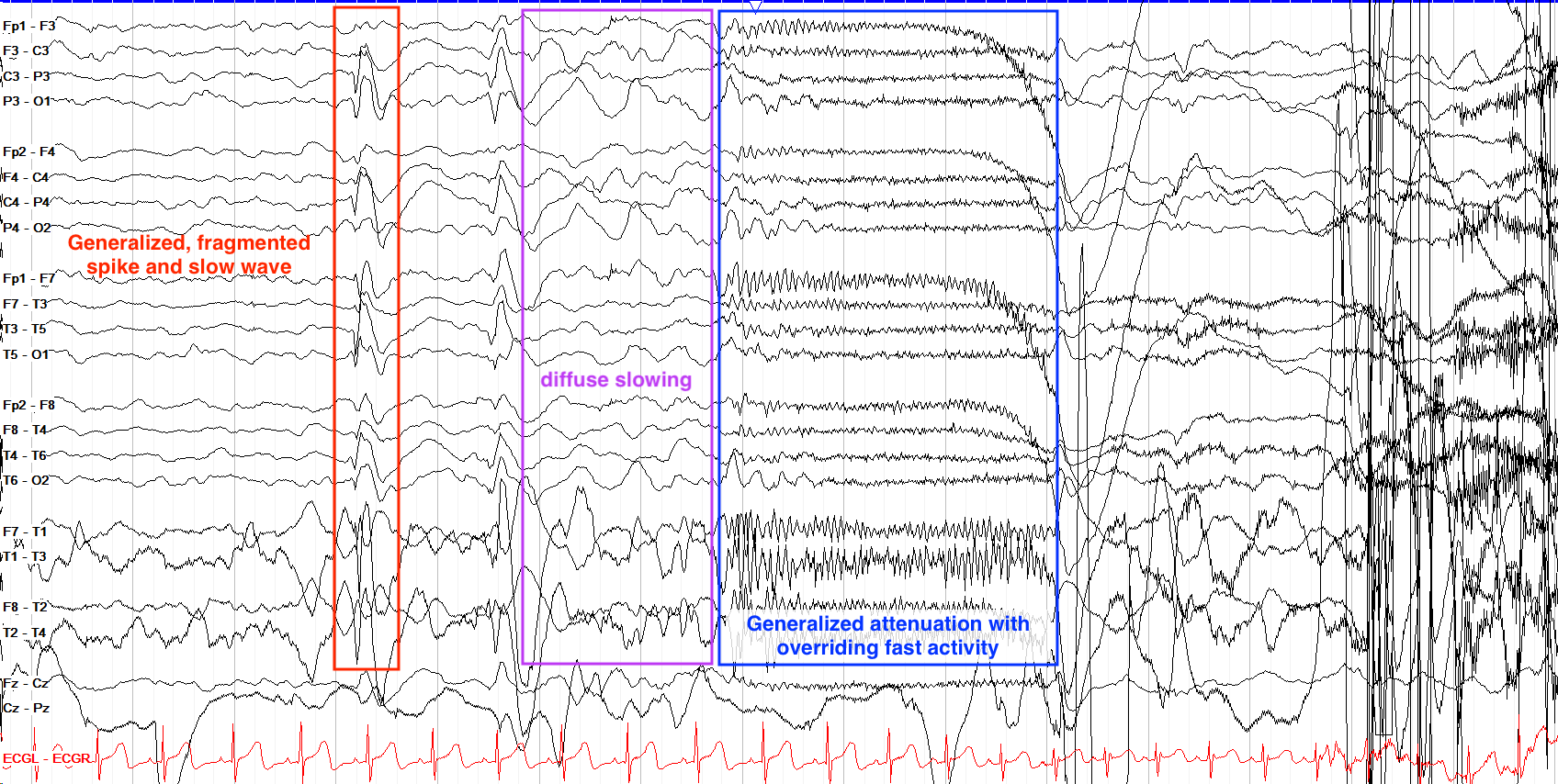
Tonic seizures are clinically marked by tonic muscle stiffening, and are mostly generalized; they do not evolve into clonic activity. Their appearance on EEG can look somewhat like artifact to the untrained eye, and is classically comprised of an early diffuse overriding slow wave followed by a diffuse electrodecrement with superimposed fast activity which then builds back up into slightly higher but still relatively low amplitude diffuse fast activity, at times with spikes and sharps.
Many tonic seizures are generalized, but on the example below note the initial sharp in the left temporal region (a “herald spike” some may call it), leading into subtle attenuation and left temporal fast activity that quickly evolves into a generalized slow wave with overriding very fast activity, then generalized tonic activity marked by myogenic artifact and fast activity.
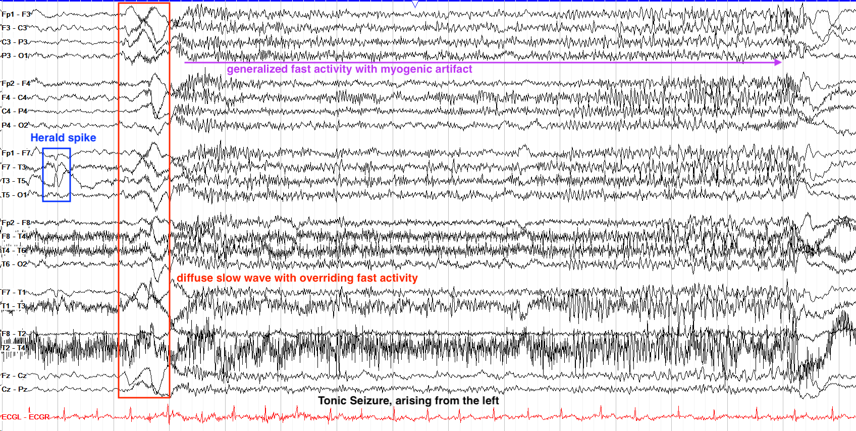
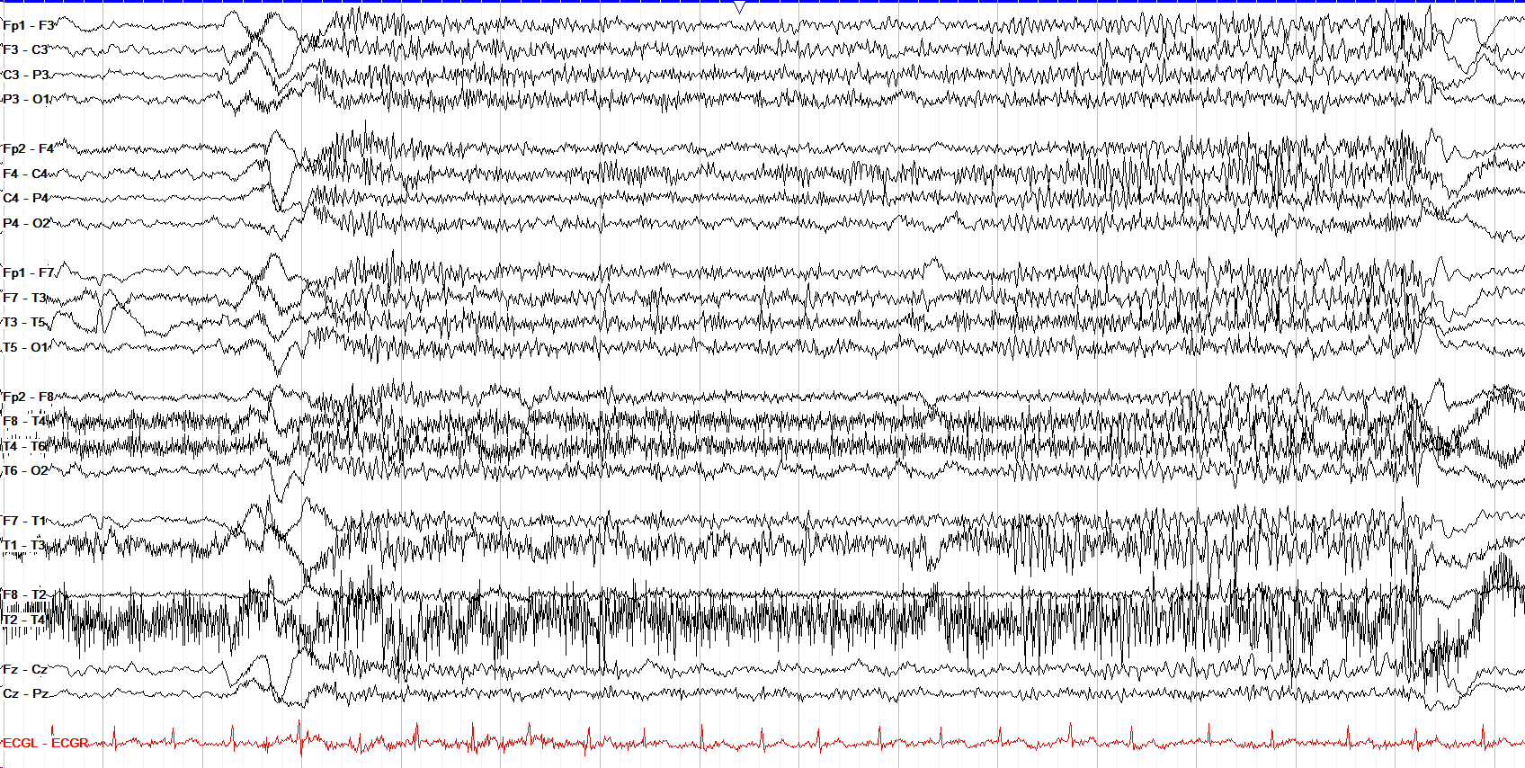
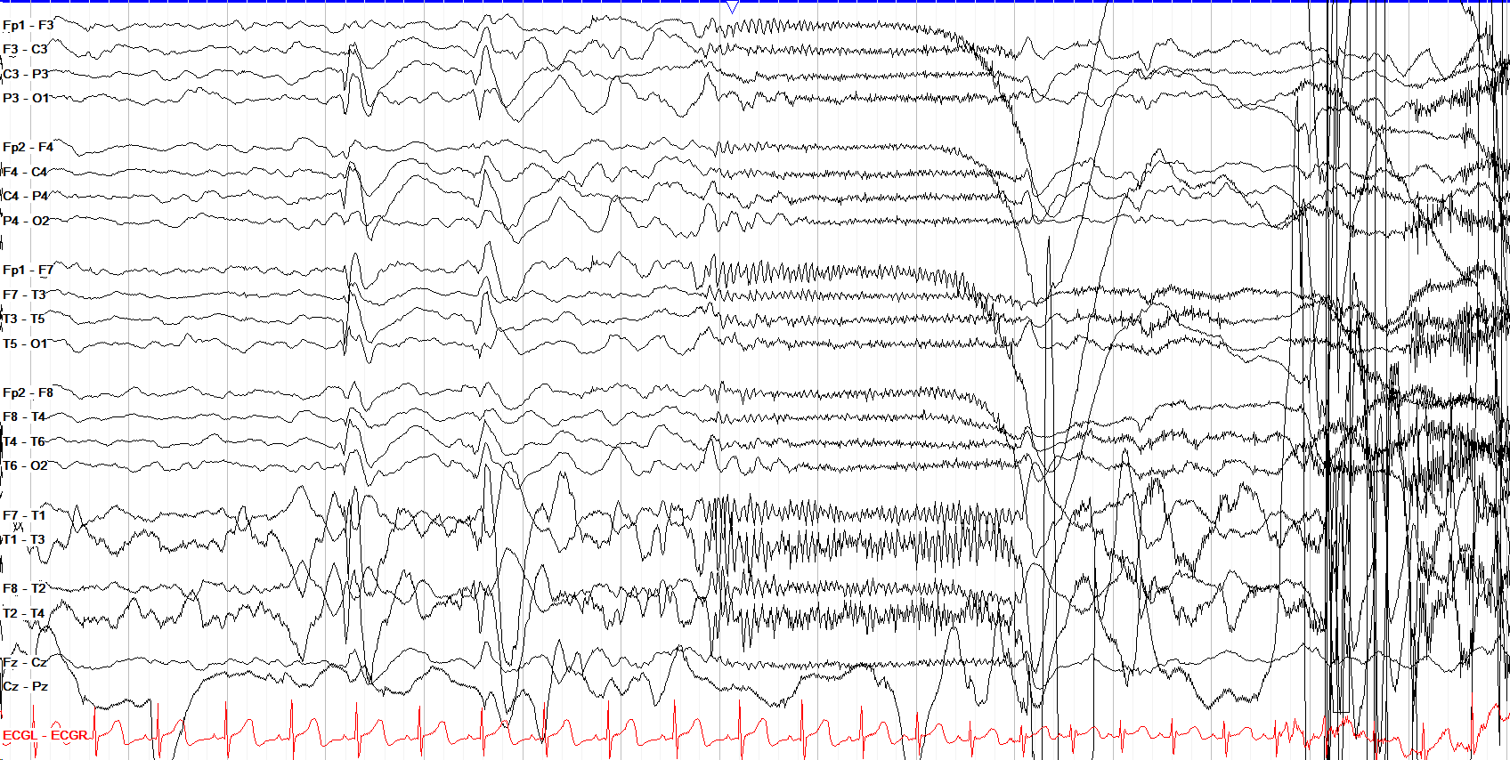
This seizure begins with a generalized albeit somewhat fragmented spike and wave discharge (note that the discharge is seen with similar morphology in all the chains), with subsequent slowing for several seconds before onset of generalized attenuation with overriding fast activity, the hallmark appearance of a tonic seizure.
Myoclonic Seizures
Myoclonus describes a brief, involuntary, shock-like jerk. Not all myoclonus is a seizure, as it can come from the cortex, subcortical regions, or spinal cord, and an occasional jerk without EEG correlate while falling asleep is normal, termed a hypnic jerk or hypnagogic myoclonus.
Myoclonus becomes a myoclonic seizure when it has a clear, time-locked epileptiform discharge (usually a generalized spike or polyspike and wave) associated with the movement on EEG. Even a single jerk with a single associated discharge is considered a myoclonic seizure (an exception to the usual requirement that seizures evolve and last ten seconds or more).
Because myoclonic seizures can be so brief, they can be easy to pass up as just interictal activity if there is no visible or documented clinical correlate. In the example below, for instance, the noted polyspikes are seen at various points in this patient’s tracing, but in this instance there was a clinical jerk to correlate. Myoclonic seizures can also be missed amongst their associated myogenic artifact, so careful inspection of the tracing is necessary.
The two most common scenarios in which you’ll see myoclonus are likely juvenile myoclonic epilepsy (discussed in the Pediatric section, and commonly associated with morning-predominant myoclonic jerks and generalized tonic-clonic seizures), and anoxic brain injury.
Anoxic injury, however, can lead to two very different types of myoclonus with two very different prognoses, the differentiation of which largely depends on their EEG. When myoclonus begins acutely after anoxia, in the first few hours when a patient is comatose, and the EEG shows generalized epileptiform activity with a severely slow (often burst suppressed) background, the prognosis is grimmer and if the discharges and correlated jerks are sustained that is considered myoclonic status epilepticus (discussed below). When the myoclonus arises only days or weeks after the anoxic injury when a patient is conscious again, and the jerks arise more due to movement and stimulation, that’s termed Lance-Adams syndrome and has a better prognosis. These two extremes—acute myoclonic seizures / status and Lance-Adams syndrome—can overlap at onset, as well, making early prognosis after anoxic injury more nebulous than long thought.
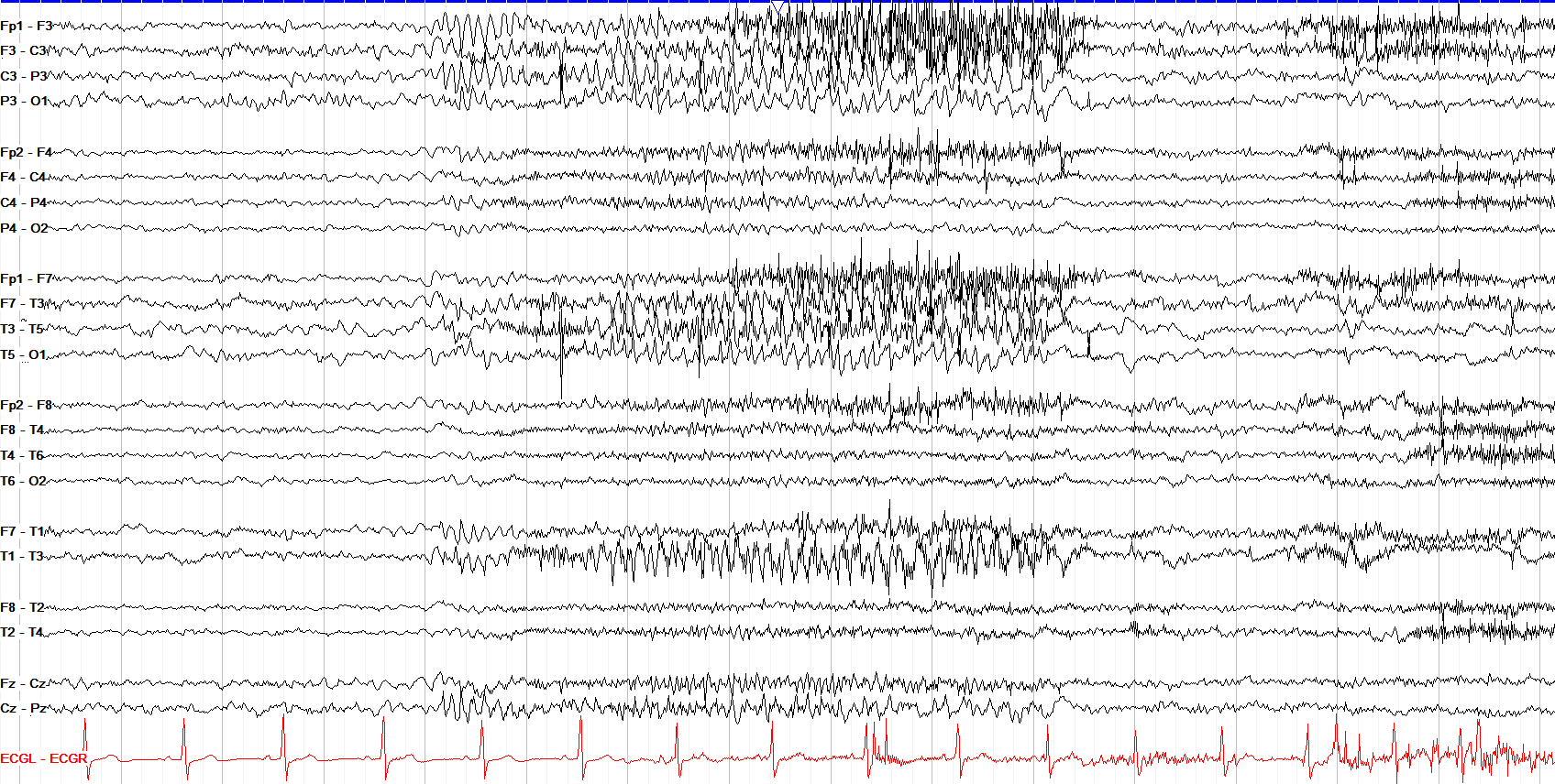
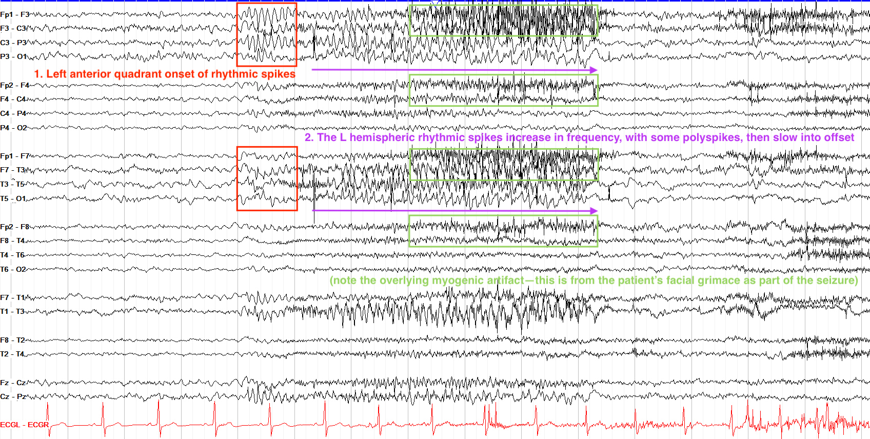
This seizure starts with a second of rhythmic spikes best seen over the left frontocentral regions, but with simultaneous albeit less well formed spikes over the left anterior temporal region. With the frontal regions of both the parasagittal and temporal chains being involved, you may see this termed as a left anterior quadrant onset seizure. Note that this one continues to evolve via higher amplitude spikes that increase in frequency and amplitude for several seconds with overriding frontally predominant myogenic artifact, then slow prior to offset. It remains focal to the left hemisphere, but there's also associated muscle artifact over the right hemisphere.
Absence Seizures
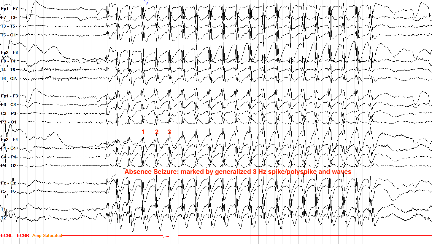
Absence seizures are a type of generalized seizure. While they’re also discussed in the pediatric section (as they are more commonly seen in children), they’re worth a second look because typical absence seizures are defined electrographically by a very classic 3 Hz generalized spike and wave or polyspike and wave morphology that is brief (often less than ten seconds) with fast on and offset, no clear postictal slowing of the background, and often little or no evolution (another exception to the rule that seizures require evolution and last ten or more seconds). Outside of formal absence epilepsy, absence seizures can occur with varying focal predominance, frequencies, and morphologies.
Whatever their electrographic pattern, absence seizures are marked clinically by abrupt and brief cessation of awareness and activity, often with eye fluttering or blinking, with rapid return to normal after the seizure ends. Atypical absence seizures are often a hair slower, trending at 2.5 Hz or less with less abrupt on / offset, and can come with other features such as motor activity or more prolonged postictal states; they are often seen with broader developmental and epileptic encephalopathies like Lennox-Gastaut Syndrome.
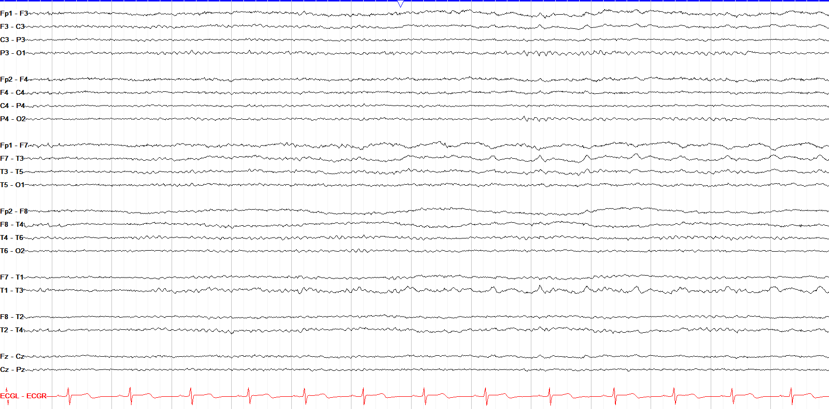
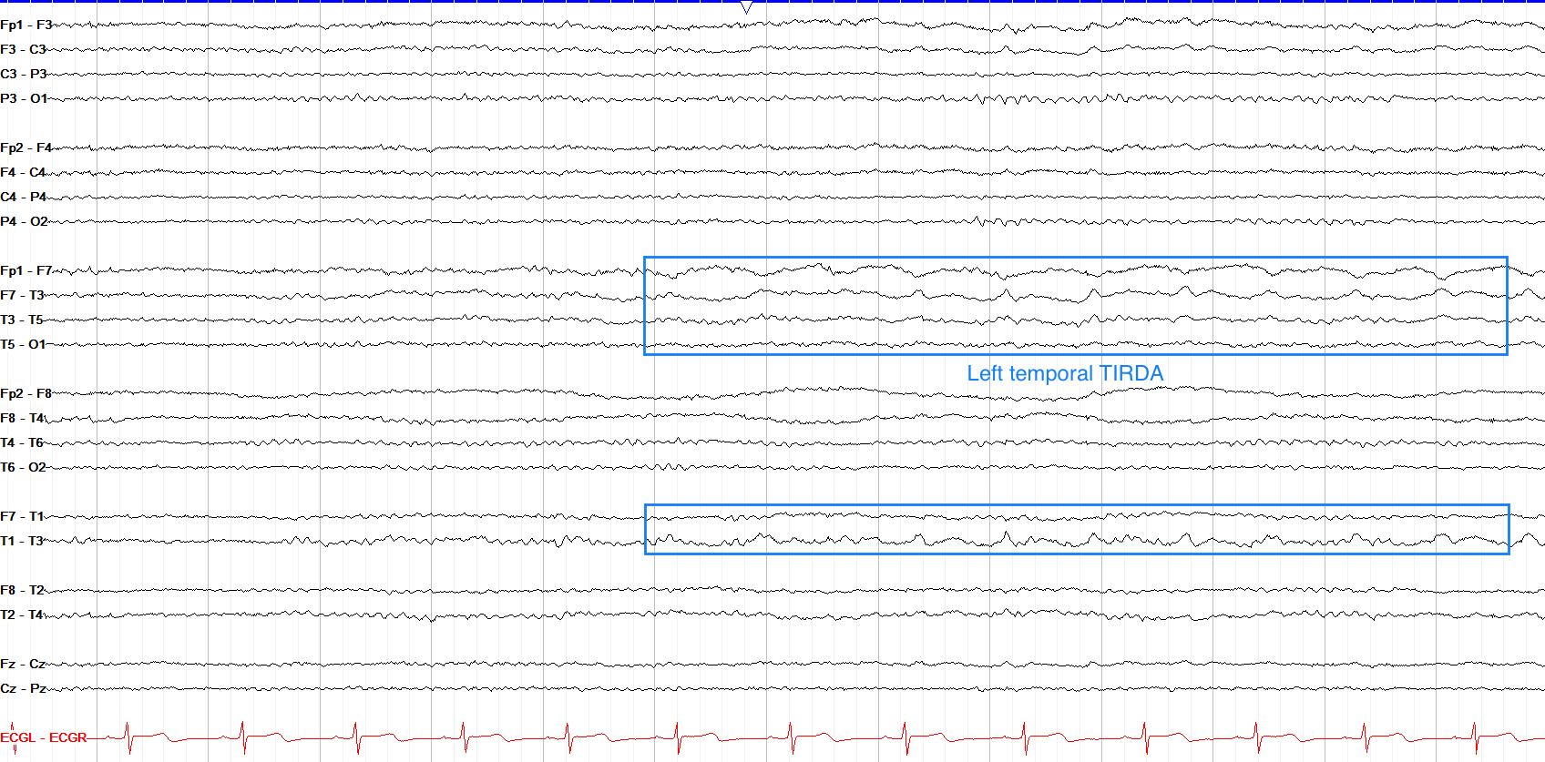
This page shows anterior left temporal sharply contoured delta activity at about 1 Hz, but there is no evolution that we can see on this page so it doesn't fit criteria for seizure. This is likely an example of temporal lateralized rhythmic delta activity (LRDA, previously termed TIRDA), which sits on the IIC as an interictal rather than overtly ictal finding. The caveat to this is that we don’t see the following page; if there is some evolution in frequency or distribution of this activity, seizure would return as a possibility.
Spasms
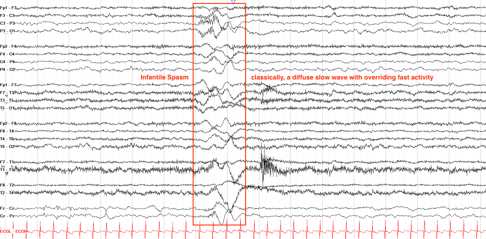
Spasms are most commonly seen in the setting of infantile spasms, or infantile epileptic spasms syndrome (IESS, previously termed West Syndrome), and thus come along with the hypsarrhythmia background of high amplitude, very disorganized activity with multifocal spikes. However, spasms can also be present in other epilepsies such as Lennox-Gastaut.
Electrographically, spasms are an unusual seizure type, classically appearing as brief and diffuse, high amplitude slow waves followed by a diffuse electrodecrement, often with superimposed fast activity. Their associated clinical correlate, usually a head drop with bilateral tonic arm raise and extension, can lead to significant overlying movement and myogenic artifact.
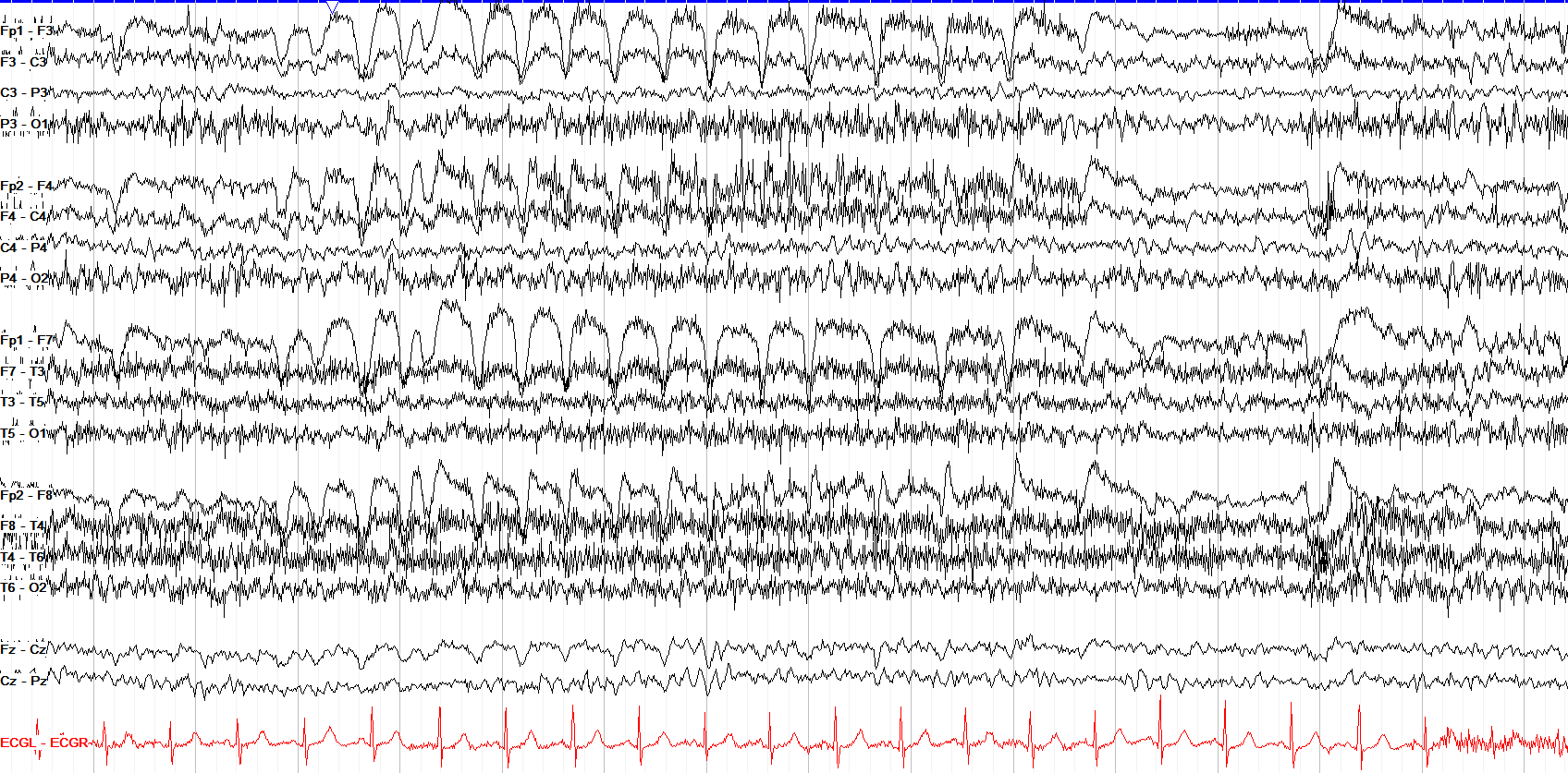
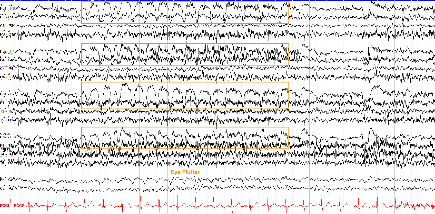
This tracing shows about seven seconds of bifrontal rhythmic activity. Note, however, that while there might appear to be evolution here, the change is very localized to the frontal leads, and their morphology is not consistent with typical epileptiform spike wave activity. This is actually a pretty classic example of eye flutter.
Also remember that seizures require at least ten seconds of evolution if there is no clinical correlate, so even if this were epileptiform in nature, it still wouldn't be categorized as a seizure unless there was a clinical correlate.
Status Epilepticus
While most seizures stop on their own within a few minutes or less, those that do not are termed status epilepticus (SE). This can arise due to either failure of the normal inhibitory systems that prevent and stop seizures, or activation of systems that pathologically propagate the seizure.
Per the International League Against Epilepsy (ILAE) there are two main time points to focus on for status epilepticus; namely, t1 is the point past which a seizure is unlikely to stop by itself and thus requires emergent abortive treatment, and t2 is the point past which ongoing seizure activity leads to brain injury, altered network function and connection, and potential deficit.
While not all seizure types have clear measured times for these values, some do and those timepoints are important to know as you read EEGs that show ongoing seizure activity:
| Seizure type | t1 | t2 |
|---|---|---|
| Tonic clonic | 5 min | 30 min |
| Focal, with impaired consciousness | 10 min | >60 min |
| Absence | 10–15 min | not established |
| Tonic, myoclonic, spasms | not established | not established |
The types without an established threshold require clinical judgment, but generally speaking seizures should be treated as promptly as is safely possible. Of note, post-anoxic myoclonic status epilepticus, while without a clear t1 / t2, is commonly defined as repetitive jerks with EEG correlate continuing beyond thirty minutes.
While many seizures have a clinical correlate, when status epilepticus has minimal or no outward sign it’s termed non-convulsive status epilepticus (NCSE). While this can come with fluctuating mental status impairment, NCSE can only be reliably diagnosed via EEG, on which it is defined by an electrographic seizure going for ten or more continuous minutes, or involving at least 20% of an hour on the tracing even if it starts and stops intermittently. Many critically ill patients are at risk for NCSE, so one who is unresponsive or with fluctuating mental status without a clear cause should be considered for continuous EEG monitoring.
- Seizures require evolution in frequency, morphology, or distribution, and should last ten or more seconds unless there is a clear clinical correlate (then they can be shorter).
- Generalized seizures do not have a clear location of onset and usually involve impaired consciousness, but do not necessitate tonic clonic activity.
- Focal seizures arise from a particular region of the brain, and can come with or without impaired consciousness.
- Focal seizures can evolve to bilateral tonic clonic seizures, and it’s important to differentiate these from generalized tonic clonic seizures because appropriate medications for each type differ.
- Tonic seizures are classically marked by a large slow wave followed by diffuse electrodecrement with overriding fast activity.
- Myoclonic seizures often don’t show evolution, are very brief (even less than a second), and are characterized by usually generalized discharges with an associated clinical jerk.
- Typical absence seizures arise with 3 Hz generalized spike and wave activity, and are very brief without a postictal state.
- Spasms are most commonly seen in infants, and have a high amplitude slow wave leading into diffuse electrodecrement with superimposed fast activity.
- Status epilepticus (SE) describes seizure activity that doesn’t stop on its own, and can occur with all seizure types; non-convulsive status epilepticus (NCSE) has minimal or no outward signs and is only reliably seen by EEG.